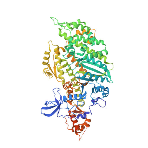
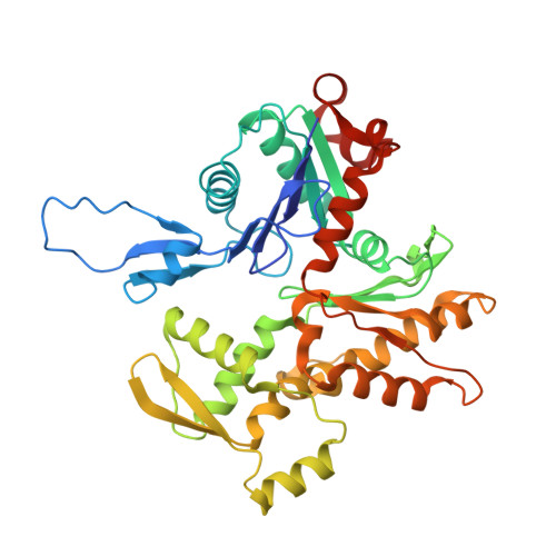
8EFH

Helical reconstruction of the human cardiac actin-tropomyosin-myosin complex in complex with ADP-Mg2+


Experimental Data Snapshot

  • Method: ELECTRON MICROSCOPY
  • Resolution: 3.30 Å
  • Aggregation State: FILAMENT 
  • Reconstruction Method: HELICAL 

wwPDB Validation   3D Report Full Report


This is version 1.1 of the entry. See complete history


Literature

Conformational changes linked to ADP release from human cardiac myosin bound to actin-tropomyosin.

Doran, M.H.Rynkiewicz, M.J.Rassici, D.Bodt, S.M.L.Barry, M.E.Bullitt, E.Yengo, C.M.Moore, J.R.Lehman, W.

(2023) J Gen Physiol 155

  • DOI: https://doi.org/10.1085/jgp.202213267
  • Primary Citation of Related Structures:  
    8EFD, 8EFE, 8EFH

  • PubMed Abstract: 

    Following binding to the thin filament, β-cardiac myosin couples ATP-hydrolysis to conformational rearrangements in the myosin motor that drive myofilament sliding and cardiac ventricular contraction. However, key features of the cardiac-specific actin-myosin interaction remain uncertain, including the structural effect of ADP release from myosin, which is rate-limiting during force generation. In fact, ADP release slows under experimental load or in the intact heart due to the afterload, thereby adjusting cardiac muscle power output to meet physiological demands. To further elucidate the structural basis of this fundamental process, we used a combination of cryo-EM reconstruction methodologies to determine structures of the human cardiac actin-myosin-tropomyosin filament complex at better than 3.4 Å-resolution in the presence and in the absence of Mg2+·ADP. Focused refinements of the myosin motor head and its essential light chains in these reconstructions reveal that small changes in the nucleotide-binding site are coupled to significant rigid body movements of the myosin converter domain and a 16-degree lever arm swing. Our structures provide a mechanistic framework to understand the effect of ADP binding and release on human cardiac β-myosin, and offer insights into the force-sensing mechanism displayed by the cardiac myosin motor.


  • Organizational Affiliation

    School of Medicine, Department of Physiology and Biophysics, Boston University , Boston, MA, USA.


Macromolecules
Find similar proteins by:  (by identity cutoff)  |  3D Structure
Entity ID: 1
MoleculeChains Sequence LengthOrganismDetailsImage
beta-cardiac myosin II865Homo sapiensMutation(s): 0 
Gene Names: MYH7MYHCB
UniProt & NIH Common Fund Data Resources
Find proteins for P12883 (Homo sapiens)
Explore P12883 
Go to UniProtKB:  P12883
PHAROS:  P12883
GTEx:  ENSG00000092054 
Entity Groups  
Sequence Clusters30% Identity50% Identity70% Identity90% Identity95% Identity100% Identity
UniProt GroupP12883
Sequence Annotations
Expand
  • Reference Sequence
Find similar proteins by:  (by identity cutoff)  |  3D Structure
Entity ID: 2
MoleculeChains Sequence LengthOrganismDetailsImage
Actin, alpha cardiac muscle 1B [auth F],
C [auth B],
D [auth C],
E [auth D],
F [auth E]
377Sus scrofaMutation(s): 0 
UniProt
Find proteins for P68137 (Sus scrofa)
Explore P68137 
Go to UniProtKB:  P68137
Entity Groups  
Sequence Clusters30% Identity50% Identity70% Identity90% Identity95% Identity100% Identity
UniProt GroupP68137
Sequence Annotations
Expand
  • Reference Sequence
Find similar proteins by:  (by identity cutoff)  |  3D Structure
Entity ID: 3
MoleculeChains Sequence LengthOrganismDetailsImage
Tropomyosin alpha-1 chainG [auth O],
H [auth P]
284Homo sapiensMutation(s): 0 
Gene Names: TPM1C15orf13TMSA
UniProt & NIH Common Fund Data Resources
Find proteins for P09493 (Homo sapiens)
Explore P09493 
Go to UniProtKB:  P09493
PHAROS:  P09493
GTEx:  ENSG00000140416 
Entity Groups  
Sequence Clusters30% Identity50% Identity70% Identity90% Identity95% Identity100% Identity
UniProt GroupP09493
Sequence Annotations
Expand
  • Reference Sequence
Small Molecules
Ligands 2 Unique
IDChains Name / Formula / InChI Key2D Diagram3D Interactions
ADP (Subject of Investigation/LOI)
Query on ADP

Download Ideal Coordinates CCD File 
J [auth A]
K [auth F]
M [auth B]
O [auth C]
Q [auth D]
J [auth A],
K [auth F],
M [auth B],
O [auth C],
Q [auth D],
S [auth E]
ADENOSINE-5'-DIPHOSPHATE
C10 H15 N5 O10 P2
XTWYTFMLZFPYCI-KQYNXXCUSA-N
MG (Subject of Investigation/LOI)
Query on MG

Download Ideal Coordinates CCD File 
I [auth A]
L [auth F]
N [auth B]
P [auth C]
R [auth D]
I [auth A],
L [auth F],
N [auth B],
P [auth C],
R [auth D],
T [auth E]
MAGNESIUM ION
Mg
JLVVSXFLKOJNIY-UHFFFAOYSA-N
Experimental Data & Validation

Experimental Data

  • Method: ELECTRON MICROSCOPY
  • Resolution: 3.30 Å
  • Aggregation State: FILAMENT 
  • Reconstruction Method: HELICAL 
EM Software:
TaskSoftware PackageVersion
MODEL REFINEMENTPHENIX1.18
RECONSTRUCTIONRELION

Structure Validation

View Full Validation Report


Entry History & Funding Information

Deposition Data


Funding OrganizationLocationGrant Number
National Institutes of Health/National Heart, Lung, and Blood Institute (NIH/NHLBI)United StatesR01HL036153

Revision History  (Full details and data files)

  • Version 1.0: 2023-01-11
    Type: Initial release
  • Version 1.1: 2023-01-25
    Changes: Database references