8DWE | pdb_00008dwe

Adenine glycosylase MutY variant E43Q in complex with DNA containing d(8-oxo-G) paired with substrate purine


Experimental Data Snapshot

  • Method: X-RAY DIFFRACTION
  • Resolution: 2.20 Å
  • R-Value Free: 
    0.234 (Depositor), 0.234 (DCC) 
  • R-Value Work: 
    0.192 (Depositor), 0.192 (DCC) 
  • R-Value Observed: 
    0.193 (Depositor) 

Starting Model: experimental
View more details

wwPDB Validation 3D Report Full Report

Validation slider image for 8DWE

This is version 1.5 of the entry. See complete history

Literature

Structural Basis for Nucleobase Activation by the Adenine DNA Glycosylase MutY.

Russelburg, L.P.Demir, M.Cedeno, K.David, S.S.Horvath, M.P.

(2026) Chembiochem 27: e70414-e70414

  • DOI: https://doi.org/10.1002/cbic.70414
  • Primary Citation Related Structures: 
    8DWD, 8DWE, 8DWF

  • PubMed Abstract: 

    The DNA glycosylase MutY excises adenine when mispaired with oxidized guanine (OG). While it is understood that inappropriate adenine excision would be catastrophic, the mechanism by which MutY activity is kept in check and only licensed at OG:A lesions is unknown. To explore the structural basis for nucleobase activation, we tested kinetic and structural consequences following replacement of the catalytic Glu, a signature residue for MutY. E43Q and E43S substitution variants of MutY from Geobacillus stearothermophilus, though severely impaired, retained measurable activity. X-ray crystal structures showed the substrate nucleobase in an anti conformation, rotated by 180° from the syn conformation seen in previous substrate complexes. Remarkably, the AP product was observed as the alpha-anomer configuration when generated by these Glu-replacement variants, completely different from the beta-anomer AP product expected for the wild-type enzyme and seen directly for other cancer-associated variants. Our results suggest a mechanism for regulating MutY, whereby Glu engagement with the syn conformation of the nucleobase licenses a "go ahead" state for adenine excision only at OG:A lesions, while Glu dis-engagement establishes an "on hold" state to avoid inappropriate activity elsewhere.


  • Organizational Affiliation
    • School of Biological Sciences, University of Utah, Salt Lake City, Utah, USA.

Macromolecule Content 

  • Total Structure Weight: 98.24 kDa 
  • Atom Count: 6,611 
  • Modeled Residue Count: 751 
  • Deposited Residue Count: 774 
  • Unique protein chains: 1
  • Unique nucleic acid chains: 2

Macromolecules


Find similar proteins by:|  3D Structure
Entity ID: 1
MoleculeChains  Sequence LengthOrganismDetailsImage
Adenine DNA glycosylase
A, D
365Geobacillus stearothermophilusMutation(s): 1 
Gene Names: mutY
EC: 3.2.2.31
UniProt
Find proteins for P83847 (Geobacillus stearothermophilus)
Explore P83847 
Go to UniProtKB:  P83847
Entity Groups
Sequence Clusters30% Identity50% Identity70% Identity90% Identity95% Identity100% Identity
UniProt GroupP83847
Sequence Annotations
Expand
Reference Sequence
Find similar nucleic acids by:  Sequence
Entity ID: 2
MoleculeChains LengthOrganismImage
DNA (5'-D(*AP*AP*GP*AP*CP*(8OG)P*TP*GP*GP*AP*C)-3')
B, E
11synthetic construct
Sequence Annotations
Expand
Reference Sequence
Find similar nucleic acids by:  Sequence
Entity ID: 3
MoleculeChains LengthOrganismImage
DNA (5'-D(*TP*GP*TP*CP*CP*AP*(PRN)P*GP*TP*CP*T)-3')
C, F
11synthetic construct
Sequence Annotations
Expand
Reference Sequence

Small Molecules

Ligands 4 Unique
IDChains Name / Formula / InChI Key2D Diagram3D Interactions
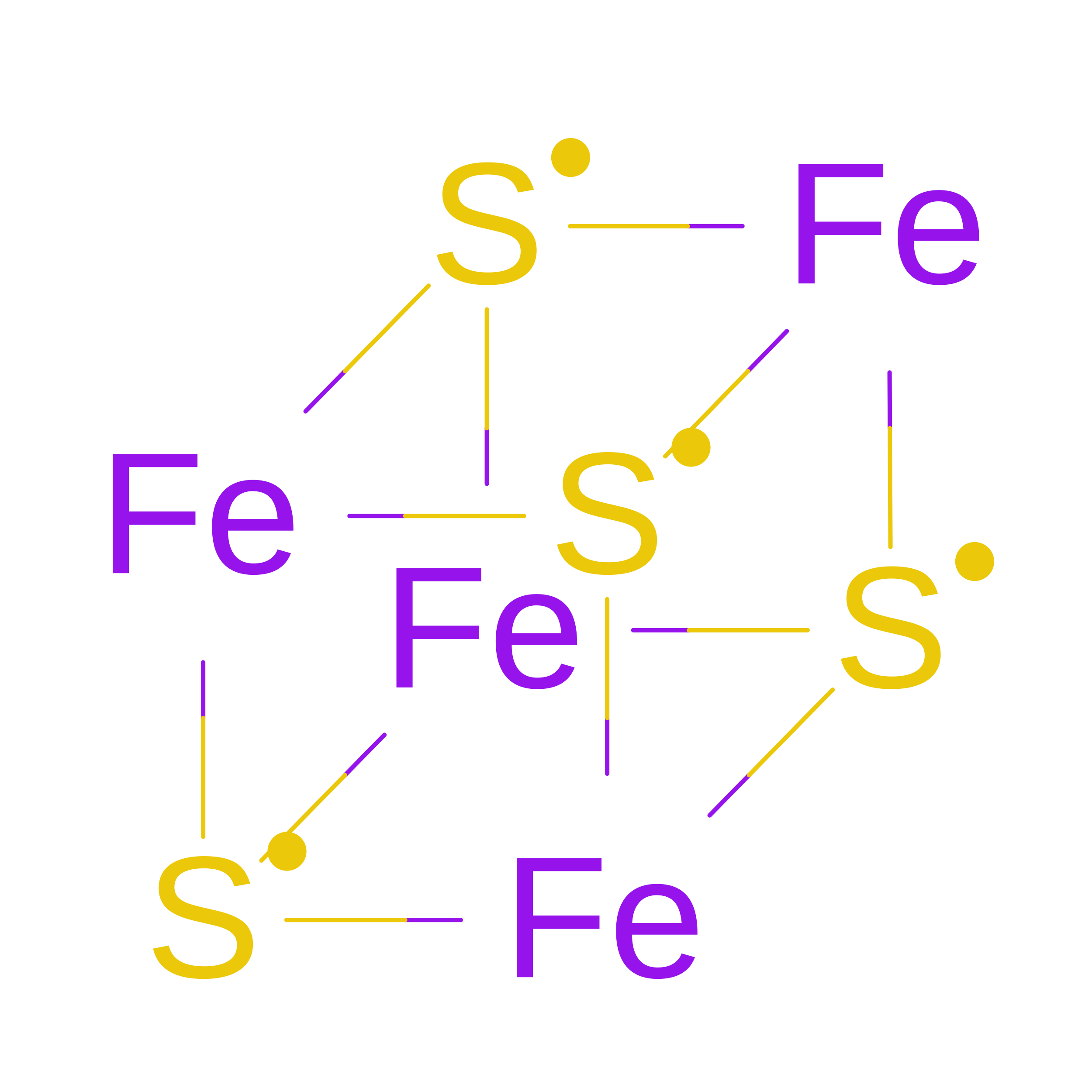
SF4
(Subject of Investigation/LOI)

Query on SF4


Download:Ideal Coordinates CCD File
G [auth A],
L [auth D]
IRON/SULFUR CLUSTER
Fe4 S4
LJBDFODJNLIPKO-UHFFFAOYSA-N
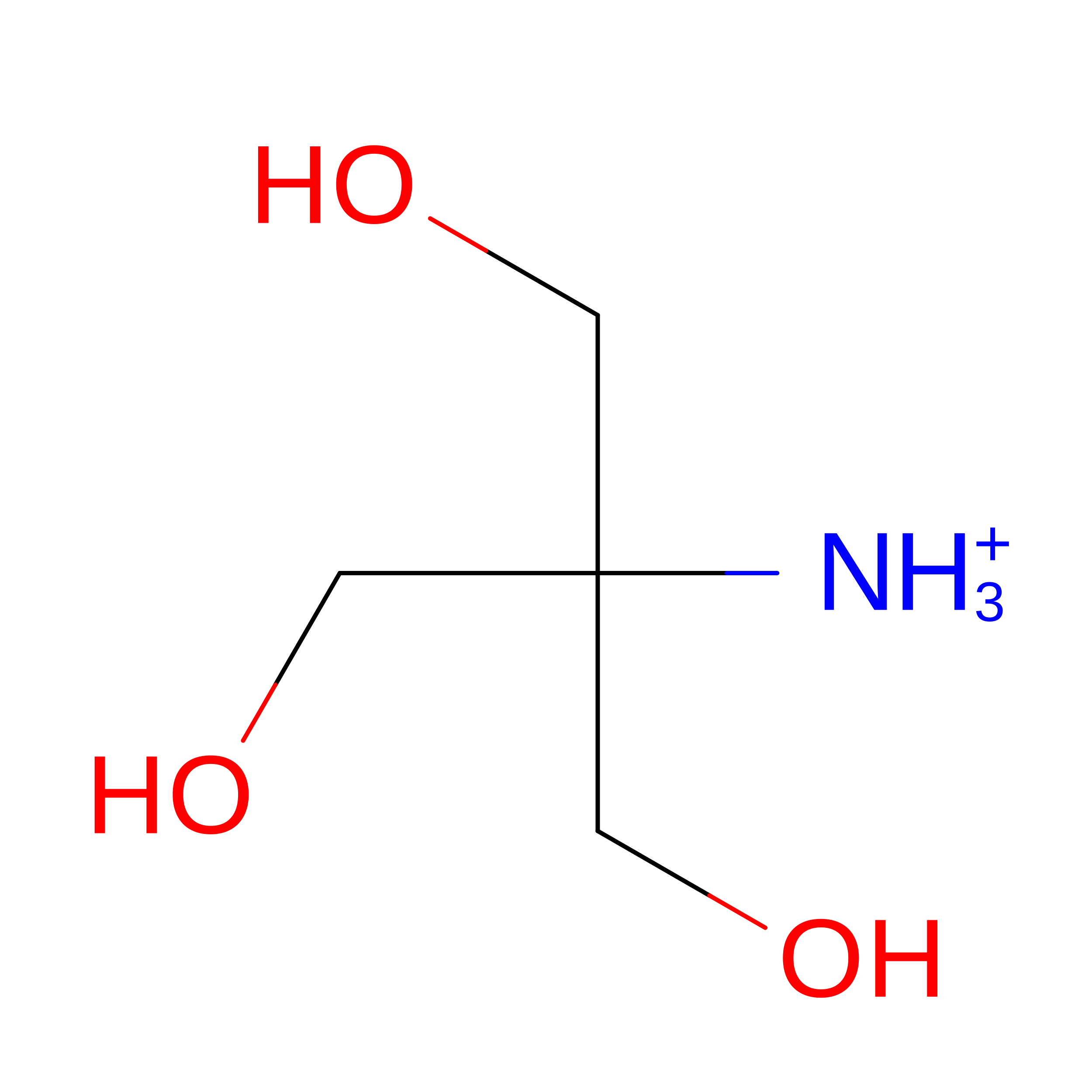
TRS

Query on TRS


Download:Ideal Coordinates CCD File
K [auth B],
P [auth E]
2-AMINO-2-HYDROXYMETHYL-PROPANE-1,3-DIOL
C4 H12 N O3
LENZDBCJOHFCAS-UHFFFAOYSA-O
EDO

Query on EDO


Download:Ideal Coordinates CCD File
H [auth A],
M [auth D]
1,2-ETHANEDIOL
C2 H6 O2
LYCAIKOWRPUZTN-UHFFFAOYSA-N
CA

Query on CA


Download:Ideal Coordinates CCD File
I [auth A],
J [auth A],
N [auth D],
O [auth D]
CALCIUM ION
Ca
BHPQYMZQTOCNFJ-UHFFFAOYSA-N

Experimental Data & Validation

Experimental Data

  • Method: X-RAY DIFFRACTION
  • Resolution: 2.20 Å
  • R-Value Free:  0.234 (Depositor), 0.234 (DCC) 
  • R-Value Work:  0.192 (Depositor), 0.192 (DCC) 
  • R-Value Observed: 0.193 (Depositor) 
Space Group: P 1 21 1
Unit Cell:
Length ( Å )Angle ( ˚ )
a = 48.63α = 90
b = 137.96β = 101
c = 74.49γ = 90
Software Package:
Software NamePurpose
PHENIXrefinement
XDSdata reduction
XSCALEdata scaling
PHENIXphasing

Structure Validation

View Full Validation Report


Entry History 

& Funding Information

Deposition Data


Funding OrganizationLocationGrant Number
National Science Foundation (NSF, United States)United States1905304
National Science Foundation (NSF, United States)United States1905249
National Science Foundation (NSF, United States)United States1610721
National Science Foundation (NSF, United States)United States1608934

Revision History  (Full details and data files)

  • Version 1.0: 2023-08-09
    Type: Initial release
  • Version 1.1: 2024-05-22
    Changes: Data collection
  • Version 1.2: 2026-03-04
    Changes: Refinement description, Structure summary
  • Version 1.3: 2026-06-03
    Changes: Database references
  • Version 1.4: 2026-08-12
    Type: Remediation
    Reason: Metalloprotein remediation
    Changes: Derived calculations, Structure summary
  • Version 1.5: 2026-09-09
    Changes: Database references