8DVX | pdb_00008dvx

Structure of acetylated Pig somatic Cytochrome c (Aly39) at 1.5A


Experimental Data Snapshot

  • Method: X-RAY DIFFRACTION
  • Resolution: 1.50 Å
  • R-Value Free: 
    0.204 (Depositor), 0.207 (DCC) 
  • R-Value Work: 
    0.171 (Depositor), 0.168 (DCC) 
  • R-Value Observed: 
    0.172 (Depositor) 

Starting Model: experimental
View more details

wwPDB Validation 3D Report Full Report

Validation slider image for 8DVX

This is version 1.4 of the entry. See complete history

Literature

Cytochrome c lysine acetylation regulates cellular respiration and cell death in ischemic skeletal muscle.

Morse, P.T.Perez-Mejias, G.Wan, J.Turner, A.A.Marquez, I.Kalpage, H.A.Vaishnav, A.Zurek, M.P.Huettemann, P.P.Kim, K.Arroum, T.De la Rosa, M.A.Chowdhury, D.D.Lee, I.Brunzelle, J.S.Sanderson, T.H.Malek, M.H.Meierhofer, D.Edwards, B.F.P.Diaz-Moreno, I.Huttemann, M.

(2023) Nat Commun 14: 4166-4166

  • DOI: https://doi.org/10.1038/s41467-023-39820-8
  • Primary Citation Related Structures: 
    8DVX, 8DZL

  • PubMed Abstract: 

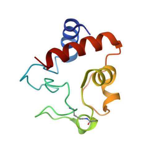
    Skeletal muscle is more resilient to ischemia-reperfusion injury than other organs. Tissue specific post-translational modifications of cytochrome c (Cytc) are involved in ischemia-reperfusion injury by regulating mitochondrial respiration and apoptosis. Here, we describe an acetylation site of Cytc, lysine 39 (K39), which was mapped in ischemic porcine skeletal muscle and removed by sirtuin5 in vitro. Using purified protein and cellular double knockout models, we show that K39 acetylation and acetylmimetic K39Q replacement increases cytochrome c oxidase (COX) activity and ROS scavenging while inhibiting apoptosis via decreased binding to Apaf-1, caspase cleavage and activity, and cardiolipin peroxidase activity. These results are discussed with X-ray crystallography structures of K39 acetylated (1.50 Å) and acetylmimetic K39Q Cytc (1.36 Å) and NMR dynamics. We propose that K39 acetylation is an adaptive response that controls electron transport chain flux, allowing skeletal muscle to meet heightened energy demand while simultaneously providing the tissue with robust resilience to ischemia-reperfusion injury.


  • Organizational Affiliation
    • Center for Molecular Medicine and Genetics, Wayne State University, Detroit, MI, 48201, USA.

Macromolecule Content 

  • Total Structure Weight: 50.5 kDa 
  • Atom Count: 3,836 
  • Modeled Residue Count: 416 
  • Deposited Residue Count: 416 
  • Unique protein chains: 1

Macromolecules

Find similar proteins by:|  3D Structure
Entity ID: 1
MoleculeChains  Sequence LengthOrganismDetailsImage
Cytochrome c
A, B, C, D
104Sus scrofaMutation(s): 0 
UniProt
Find proteins for P62895 (Sus scrofa)
Explore P62895 
Go to UniProtKB:  P62895
Entity Groups
Sequence Clusters30% Identity50% Identity70% Identity90% Identity95% Identity100% Identity
UniProt GroupP62895
Sequence Annotations
Expand
Reference Sequence

Small Molecules

Ligands 2 Unique
IDChains Name / Formula / InChI Key2D Diagram3D Interactions
HEC
(Subject of Investigation/LOI)

Query on HEC


Download:Ideal Coordinates CCD File
E [auth A],
H [auth B],
M [auth C],
O [auth D]
HEME C
C34 H34 Fe N4 O4
HXQIYSLZKNYNMH-LJNAALQVSA-N
FC6

Query on FC6


Download:Ideal Coordinates CCD File
F [auth A]
G [auth A]
I [auth B]
J [auth B]
K [auth B]
F [auth A],
G [auth A],
I [auth B],
J [auth B],
K [auth B],
L [auth B],
N [auth C]
HEXACYANOFERRATE(3-)
C6 Fe N6
HCMVSLMENOCDCK-UHFFFAOYSA-N
Modified Residues  1 Unique
IDChains TypeFormula2D DiagramParent
ALY
Query on ALY
A, B, C, D
L-PEPTIDE LINKINGC8 H16 N2 O3LYS

Experimental Data & Validation

Experimental Data

  • Method: X-RAY DIFFRACTION
  • Resolution: 1.50 Å
  • R-Value Free:  0.204 (Depositor), 0.207 (DCC) 
  • R-Value Work:  0.171 (Depositor), 0.168 (DCC) 
  • R-Value Observed: 0.172 (Depositor) 
Space Group: P 1
Unit Cell:
Length ( Å )Angle ( ˚ )
a = 34.662α = 65.97
b = 54.559β = 87.4
c = 61.782γ = 84.07
Software Package:
Software NamePurpose
PHENIXrefinement
AutoProcessdata reduction
Aimlessdata scaling
PHASERphasing

Structure Validation

View Full Validation Report


Entry History 

& Funding Information

Deposition Data


Funding OrganizationLocationGrant Number
National Institutes of Health/National Institute of General Medical Sciences (NIH/NIGMS)United States116807

Revision History  (Full details and data files)

  • Version 1.0: 2023-07-19
    Type: Initial release
  • Version 1.1: 2023-07-26
    Changes: Database references
  • Version 1.2: 2023-10-25
    Changes: Data collection, Refinement description
  • Version 1.3: 2023-11-15
    Changes: Data collection
  • Version 1.4: 2024-10-09
    Changes: Structure summary