Funding Organization(s): National Institutes of Health/National Cancer Institute (NIH/NCI), National Institutes of Health/National Institute of General Medical Sciences (NIH/NIGMS)
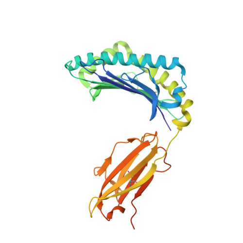
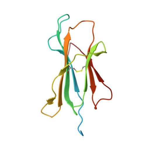
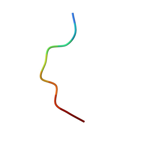
Specificity remains a major challenge to current therapeutic strategies for cancer. Mutation associated neoantigens (MANAs) are products of genetic alterations, making them highly specific therapeutic targets. MANAs are HLA-presented (pHLA) peptides derived from intracellular mutant proteins that are otherwise inaccessible to antibody-based therapeutics. Here, we describe the cryo-EM structure of an antibody-MANA pHLA complex. Specifically, we determine a TCR mimic (TCRm) antibody bound to its MANA target, the KRAS G12V peptide presented by HLA-A*03:01. Hydrophobic residues appear to account for the specificity of the mutant G12V residue. We also determine the structure of the wild-type G12 peptide bound to HLA-A*03:01, using X-ray crystallography. Based on these structures, we perform screens to validate the key residues required for peptide specificity. These experiments led us to a model for discrimination between the mutant and the wild-type peptides presented on HLA-A*03:01 based exclusively on hydrophobic interactions.
Organizational Affiliation:
Department of Biophysics and Biophysical Chemistry, The Johns Hopkins School of Medicine, Baltimore, MD, 21205, USA.
Howard Hughes Medical Institute, Chevy Chase, MD, 20815, USA.
Bloomberg~Kimmel Institute for Cancer Immunotherapy, Sidney Kimmel Comprehensive Cancer Center, Baltimore, MD, 21287, USA.
Discovery Chemistry, Protein and Structural Chemistry, Merck & Co, Inc, West Point, PA, 19846, USA.
Ludwig Center, Sidney Kimmel Comprehensive Cancer Center, Johns Hopkins University School of Medicine, Baltimore, MD, 21287, USA.
Lustgarten Pancreatic Cancer Research Laboratory, Sidney Kimmel Comprehensive Cancer Center, Johns Hopkins University School of Medicine, Baltimore, MD, 21287, USA.
Walter and Eliza Hall Institute, Parkville, VIC, 3052, Australia.
Janelia Research Campus, HHMI,19700 Helix Drive, Ashburn, VA, 20147, USA.
Energy & Photon Sciences Directorate, Brookhaven National Laboratory, Upton, NY, 11973, USA.
Case Center for Synchrotron Biosciences, Case Western Reserve University, Cleveland, OH, 44106, USA.
Novartis Institutes for BioMedical Research, 250 Massachusetts Ave, Cambridge, MA, 02139, USA.
Department of Biomedical Engineering, Johns Hopkins University, Baltimore, MD, 21218, USA.
Department of Oncology, Johns Hopkins University School of Medicine, Baltimore, MD, 21287, USA.
Division of Hematologic Malignancies and Bone Marrow Transplantation, Johns Hopkins University School of Medicine, Baltimore, MD, 21287, USA.
Division of Rheumatology, Department of Medicine, Johns Hopkins University School of Medicine, Baltimore, MD, 21224, USA.
Department of Neurosurgery, Johns Hopkins University School of Medicine, Baltimore, MD, 21205, USA.
Department of Pathology, Johns Hopkins University School of Medicine, Baltimore, MD, 21205, USA.
Bloomberg~Kimmel Institute for Cancer Immunotherapy, Sidney Kimmel Comprehensive Cancer Center, Baltimore, MD, 21287, USA. sbzhou@jhmi.edu.
Ludwig Center, Sidney Kimmel Comprehensive Cancer Center, Johns Hopkins University School of Medicine, Baltimore, MD, 21287, USA. sbzhou@jhmi.edu.
Lustgarten Pancreatic Cancer Research Laboratory, Sidney Kimmel Comprehensive Cancer Center, Johns Hopkins University School of Medicine, Baltimore, MD, 21287, USA. sbzhou@jhmi.edu.
Department of Oncology, Johns Hopkins University School of Medicine, Baltimore, MD, 21287, USA. sbzhou@jhmi.edu.
Department of Biophysics and Biophysical Chemistry, The Johns Hopkins School of Medicine, Baltimore, MD, 21205, USA. Sandra.gabelli@merck.com.
Bloomberg~Kimmel Institute for Cancer Immunotherapy, Sidney Kimmel Comprehensive Cancer Center, Baltimore, MD, 21287, USA. Sandra.gabelli@merck.com.
Department of Oncology, Johns Hopkins University School of Medicine, Baltimore, MD, 21287, USA. Sandra.gabelli@merck.com.
Department of Medicine, Johns Hopkins University School of Medicine, Baltimore, MD, 21205, USA. Sandra.gabelli@merck.com.
Discovery Chemistry, Protein and Structural Chemistry, Merck & Co, Inc, West Point, PA, 19846, USA. Sandra.gabelli@merck.com.
D [auth A] E [auth A] F [auth A] G [auth A] H [auth A]
D [auth A], E [auth A], F [auth A], G [auth A], H [auth A], I [auth A], J [auth A], L [auth B], M [auth B], N [auth B], O [auth B], P [auth B], Q [auth B]