8DJ7 | pdb_00008dj7

The complex structure between human IgG1 Fc and its high affinity receptor FcgRI H174R variant


Experimental Data Snapshot

  • Method: X-RAY DIFFRACTION
  • Resolution: 2.39 Å
  • R-Value Free: 
    0.266 (Depositor), 0.266 (DCC) 
  • R-Value Work: 
    0.214 (Depositor), 0.214 (DCC) 
  • R-Value Observed: 
    0.217 (Depositor) 

Starting Model: experimental
View more details

wwPDB Validation 3D Report Full Report

Validation slider image for 8DJ7

Ligand Structure Quality Assessment 


This is version 1.2 of the entry. See complete history

Literature

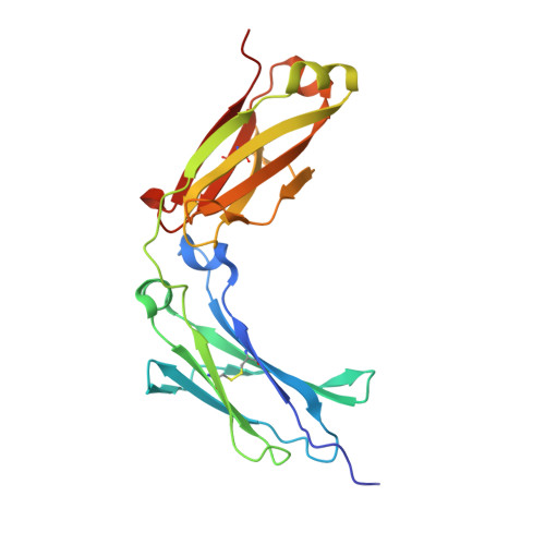
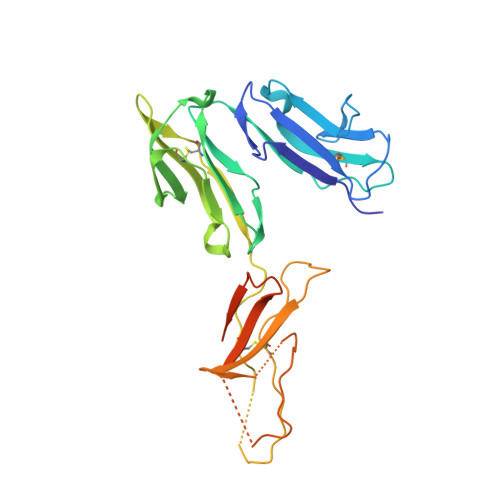
Fc gamma RI FG-loop functions as a pH sensitive switch for IgG binding and release.

Lu, J.Spencer, M.Zou, Z.Traver, M.Brzostowski, J.Sun, P.D.

(2023) Front Immunol 14: 1100499-1100499

  • DOI: https://doi.org/10.3389/fimmu.2023.1100499
  • Primary Citation Related Structures: 
    8DIN, 8DIR, 8DJ7

  • PubMed Abstract: 

    Understanding the molecular mechanism underlying the hierarchic binding between FcγRs and IgG antibodies is critical for therapeutic antibody engineering and FcγR functions. The recent determination of crystal structures of FcγRI-Fc complexes, however, resulted in two controversial mechanisms for the high affinity receptor binding to IgG. Here, we describe high resolution structures of a bovine FG-loop variant of FcγRI in complex with the Fc fragment of IgG 1 crystallized in three different conditions at neutral pH, confirming the characteristic FG loop-Fc interaction is critical to the high affinity immunoglobulin binding. We showed that the FcγRI D2-domain FG-loop functioned as a pH-sensing switch for IgG binding. Further live cell imaging of FcγRI-mediated internalization of immune complexes showed a pH sensitive temporal-spatial antibody-antigen uptake and release. Taken together, we demonstrate that the structures of FcγRI-Fc crystallized at neutral and acidic pH, respectively, represent the high and low affinity binding states of the receptor for IgG uptake and release. These results support a role for FcγRI in antigen delivery, highlight the importance of Fc glycan in antibody binding to the high affinity receptor and provide new insights to future antibody engineering.


  • Organizational Affiliation
    • Structural Immunology Section, Lab of Immunogenetics, National Institute of Allergy and Infectious Diseases, National Institutes of Health, Rockville, MD, United States.

Macromolecule Content 

  • Total Structure Weight: 83.88 kDa 
  • Atom Count: 5,663 
  • Modeled Residue Count: 669 
  • Deposited Residue Count: 715 
  • Unique protein chains: 2

Macromolecules

Find similar proteins by:|  3D Structure
Entity ID: 1
MoleculeChains  Sequence LengthOrganismDetailsImage
Ig gamma-1 Fc chainA [auth B],
B [auth A]
219Homo sapiensMutation(s): 0 
Gene Names: IGHG1
UniProt & NIH Common Fund Data Resources
Find proteins for P01857 (Homo sapiens)
Explore P01857 
Go to UniProtKB:  P01857
PHAROS:  P01857
GTEx:  ENSG00000211896 
Entity Groups
Sequence Clusters30% Identity50% Identity70% Identity90% Identity95% Identity100% Identity
UniProt GroupP01857
Glycosylation
Glycosylation Sites: 1Go to GlyGen: P01857-2
Sequence Annotations
Expand
Reference Sequence
Find similar proteins by:|  3D Structure
Entity ID: 2
MoleculeChains  Sequence LengthOrganismDetailsImage
High affinity immunoglobulin gamma Fc receptor I277Homo sapiensMutation(s): 0 
Gene Names: FCGR1AFCG1FCGR1IGFR1
UniProt & NIH Common Fund Data Resources
Find proteins for P12314 (Homo sapiens)
Explore P12314 
Go to UniProtKB:  P12314
PHAROS:  P12314
GTEx:  ENSG00000150337 
Entity Groups
Sequence Clusters30% Identity50% Identity70% Identity90% Identity95% Identity100% Identity
UniProt GroupP12314
Sequence Annotations
Expand
Reference Sequence

Oligosaccharides

Help  
Entity ID: 3
MoleculeChains Length2D Diagram GlycosylationD Interactions
2-acetamido-2-deoxy-beta-D-glucopyranose-(1-2)-alpha-D-mannopyranose-(1-3)-[2-acetamido-2-deoxy-beta-D-glucopyranose-(1-2)-alpha-D-mannopyranose-(1-6)]beta-D-mannopyranose-(1-4)-2-acetamido-2-deoxy-beta-D-glucopyranose-(1-4)-[alpha-L-fucopyranose-(1-6)]2-acetamido-2-deoxy-beta-D-glucopyranose
D
8N-Glycosylation
Glycosylation Resources
GlyTouCan: G80858MF
GlyCosmos: G80858MF
GlyGen: G80858MF
Entity ID: 4
MoleculeChains Length2D Diagram GlycosylationD Interactions
beta-D-galactopyranose-(1-4)-2-acetamido-2-deoxy-beta-D-glucopyranose-(1-2)-alpha-D-mannopyranose-(1-6)-[2-acetamido-2-deoxy-beta-D-glucopyranose-(1-2)-alpha-D-mannopyranose-(1-3)]beta-D-mannopyranose-(1-4)-2-acetamido-2-deoxy-beta-D-glucopyranose-(1-4)-[alpha-L-fucopyranose-(1-6)]2-acetamido-2-deoxy-beta-D-glucopyranose
E
9N-Glycosylation
Glycosylation Resources
GlyTouCan: G27919IH
GlyCosmos: G27919IH
GlyGen: G27919IH

Experimental Data & Validation

Experimental Data

  • Method: X-RAY DIFFRACTION
  • Resolution: 2.39 Å
  • R-Value Free:  0.266 (Depositor), 0.266 (DCC) 
  • R-Value Work:  0.214 (Depositor), 0.214 (DCC) 
  • R-Value Observed: 0.217 (Depositor) 
Space Group: C 1 2 1
Unit Cell:
Length ( Å )Angle ( ˚ )
a = 206.371α = 90
b = 89.037β = 96.81
c = 56.191γ = 90
Software Package:
Software NamePurpose
HKL-2000data reduction
HKL-2000data scaling
PHASERphasing
PHENIXrefinement
PDB_EXTRACTdata extraction

Structure Validation

View Full Validation Report


Ligand Structure Quality Assessment 


Entry History 

& Funding Information

Deposition Data


Funding OrganizationLocationGrant Number
National Institutes of Health/National Institute Of Allergy and Infectious Diseases (NIH/NIAID)United StatesDivision of ntramural research

Revision History  (Full details and data files)

  • Version 1.0: 2023-05-10
    Type: Initial release
  • Version 1.1: 2023-10-25
    Changes: Data collection, Refinement description
  • Version 1.2: 2024-11-13
    Changes: Structure summary