8DHH | pdb_00008dhh

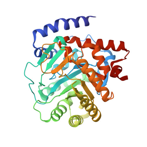
DHODH IN COMPLEX WITH LIGAND 29


Experimental Data Snapshot

  • Method: X-RAY DIFFRACTION
  • Resolution: 2.02 Å
  • R-Value Free: 
    0.216 (Depositor), 0.218 (DCC) 
  • R-Value Work: 
    0.186 (Depositor), 0.194 (DCC) 
  • R-Value Observed: 
    0.187 (Depositor) 

Starting Model: experimental
View more details

wwPDB Validation 3D Report Full Report

Validation slider image for 8DHH

Ligand Structure Quality Assessment 


This is version 1.2 of the entry. See complete history. 

Literature

N -Heterocyclic 3-Pyridyl Carboxamide Inhibitors of DHODH for the Treatment of Acute Myelogenous Leukemia.

Cisar, J.S., Pietsch, C., DeRatt, L.G., Jacoby, E., Kazmi, F., Keohane, C., Legenski, K., Matico, R., Shaffer, P., Simonnet, Y., Tanner, A., Wang, C.Y., Wang, W., Attar, R., Edwards, J.P., Kuduk, S.D.

(2022) J Med Chem 65: 11241-11256

  • DOI: https://doi.org/10.1021/acs.jmedchem.2c00788
  • Primary Citation Related Structures: 
    8DHF, 8DHG, 8DHH

  • PubMed Abstract: 

    Acute myelogenous leukemia (AML), a disease of the blood and bone marrow, is characterized by the inability of myeloblasts to differentiate into mature cell types. Dihydroorotate dehydrogenase (DHODH) is an enzyme well-known in the pyrimidine biosynthesis pathway; however, small molecule DHODH inhibitors were recently shown to induce differentiation in multiple AML subtypes. Using virtual screening and structure-based drug design approaches, a new series of N-heterocyclic 3-pyridyl carboxamide DHODH inhibitors were discovered. Two lead compounds, 19 and 29 , have potent biochemical and cellular DHODH activity, favorable physicochemical properties, and efficacy in a preclinical model of AML.


  • Organizational Affiliation: 
    • Janssen Research and Development, 1400 McKean Rd, Spring House, Pennsylvania 19477, United States.

Macromolecule Content 

  • Total Structure Weight: 41.51 kDa 
  • Atom Count: 3,193 
  • Modeled Residue Count: 358 
  • Deposited Residue Count: 369 
  • Unique protein chains: 1

Macromolecules

Find similar proteins by:|  3D Structure
Entity ID: 1
MoleculeChains  Sequence LengthOrganismDetailsImage
Dihydroorotate dehydrogenase (quinone), mitochondrial369Homo sapiensMutation(s): 0 
Gene Names: DHODH
EC: 1.3.5.2
UniProt & NIH Common Fund Data Resources
Find proteins for Q02127 (Homo sapiens)
Explore Q02127 
Go to UniProtKB:  Q02127
PHAROS:  Q02127
GTEx:  ENSG00000102967 
Entity Groups
Sequence Clusters30% Identity50% Identity70% Identity90% Identity95% Identity100% Identity
UniProt GroupQ02127
Sequence Annotations
Expand
Reference Sequence

Small Molecules

Ligands 6 Unique
IDChains Name / Formula / InChI Key2D Diagram3D Interactions
TMK
(Subject of Investigation/LOI)

Query on TMK


Download:Ideal Coordinates CCD File
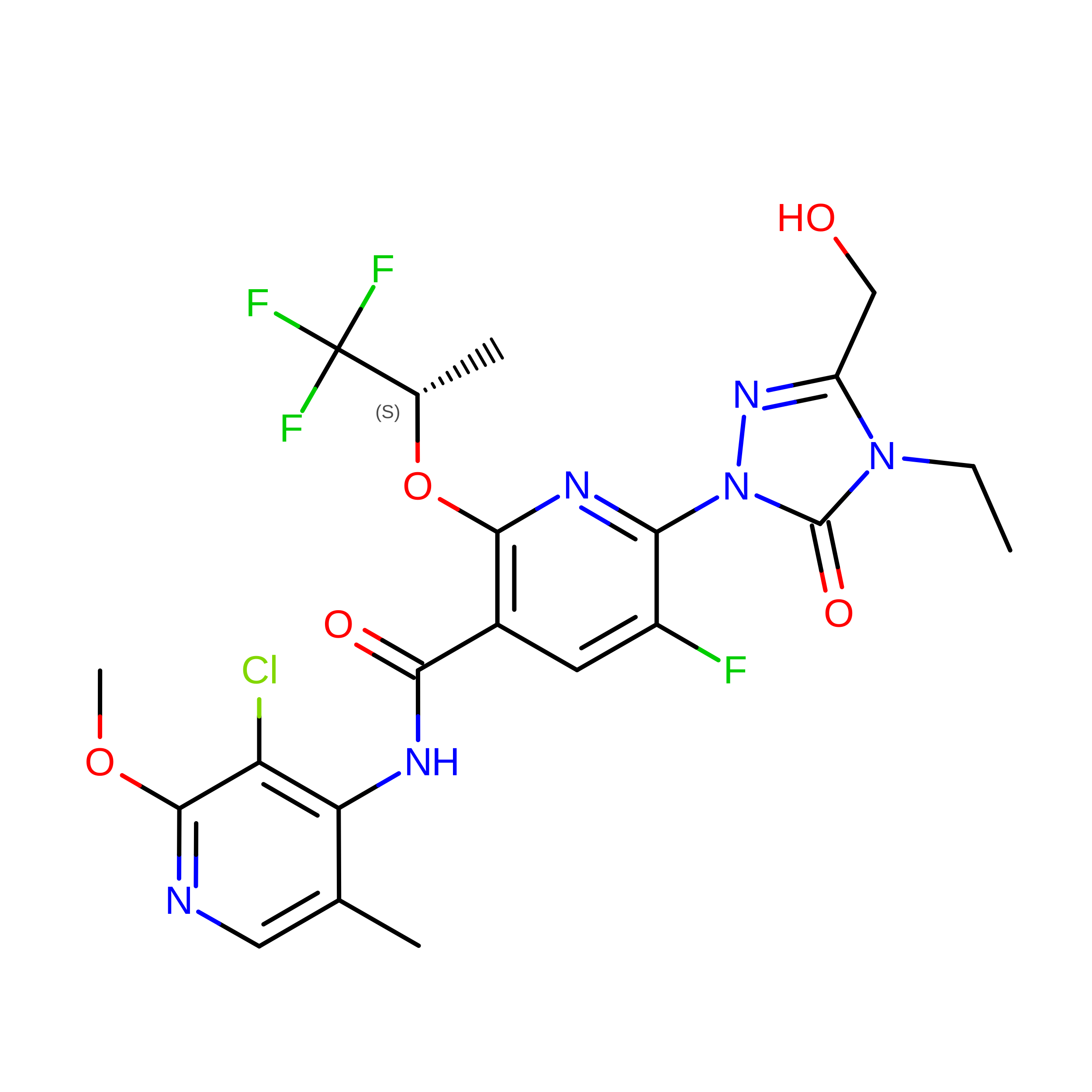
D [auth A](6M)-N-(3-chloro-2-methoxy-5-methylpyridin-4-yl)-6-[4-ethyl-3-(hydroxymethyl)-5-oxo-4,5-dihydro-1H-1,2,4-triazol-1-yl]-5-fluoro-2-{[(2S)-1,1,1-trifluoropropan-2-yl]oxy}pyridine-3-carboxamide
C21 H21 Cl F4 N6 O5
FJYNLQXYLQBCRQ-JTQLQIEISA-N
FMN

Query on FMN


Download:Ideal Coordinates CCD File
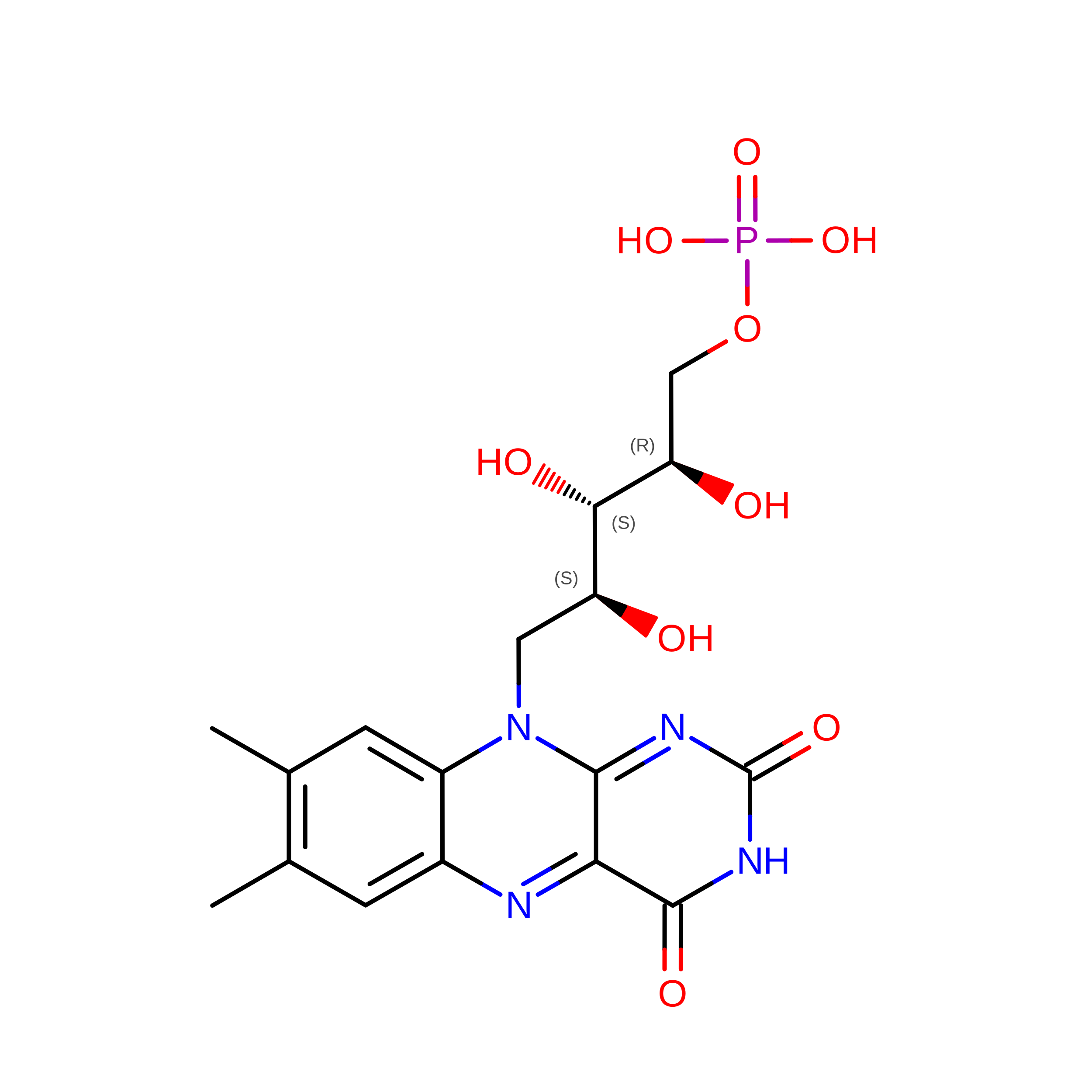
B [auth A]FLAVIN MONONUCLEOTIDE
C17 H21 N4 O9 P
FVTCRASFADXXNN-SCRDCRAPSA-N
ORO

Query on ORO


Download:Ideal Coordinates CCD File
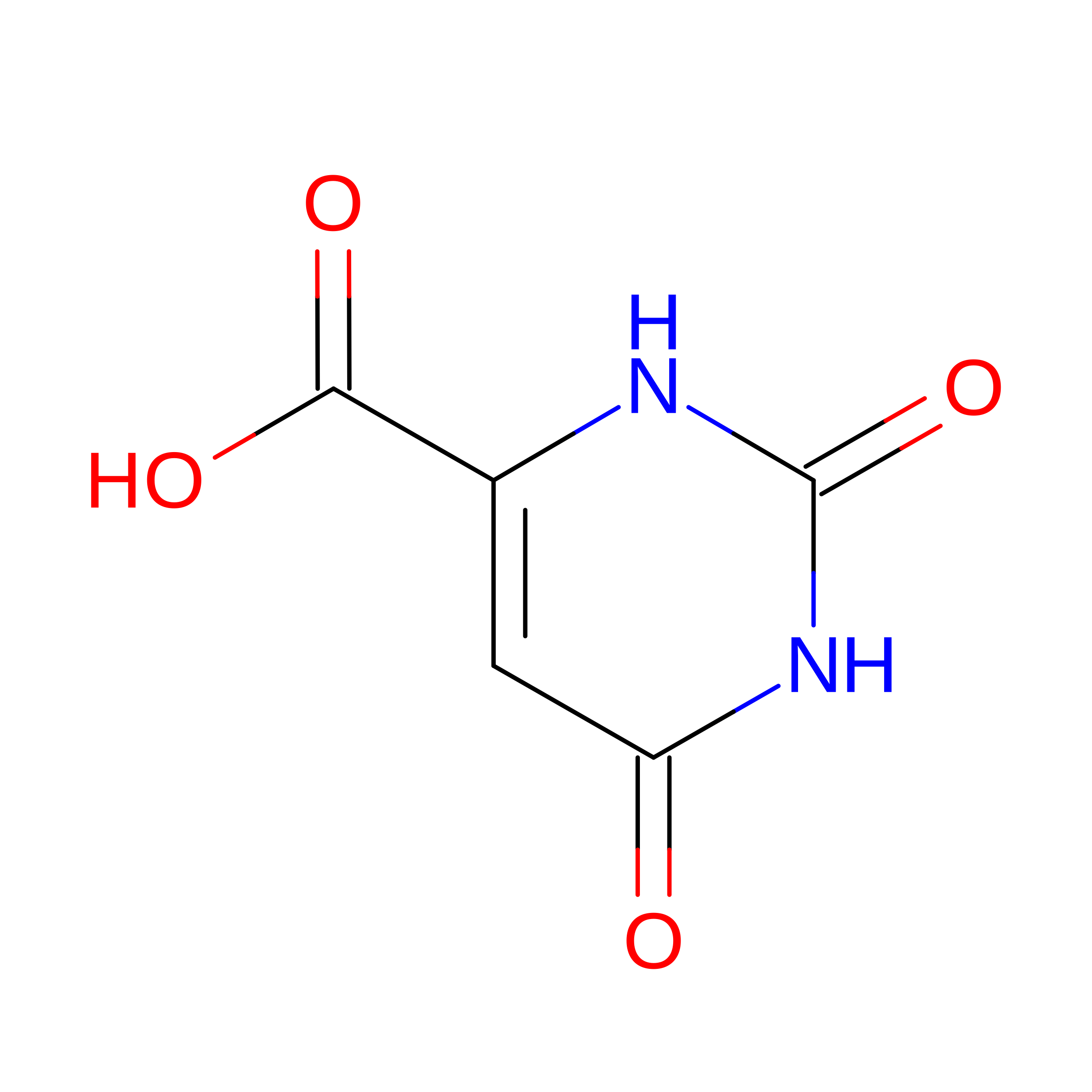
C [auth A]OROTIC ACID
C5 H4 N2 O4
PXQPEWDEAKTCGB-UHFFFAOYSA-N
SO4

Query on SO4


Download:Ideal Coordinates CCD File
I [auth A]SULFATE ION
O4 S
QAOWNCQODCNURD-UHFFFAOYSA-L
GOL

Query on GOL


Download:Ideal Coordinates CCD File
H [auth A]GLYCEROL
C3 H8 O3
PEDCQBHIVMGVHV-UHFFFAOYSA-N
ACT

Query on ACT


Download:Ideal Coordinates CCD File
E [auth A],
F [auth A],
G [auth A]
ACETATE ION
C2 H3 O2
QTBSBXVTEAMEQO-UHFFFAOYSA-M
Binding Affinity Annotations 
IDSourceBinding Affinity
TMK BindingDB:  8DHH Kd: 1.1 (nM) from 1 assay(s)
IC50: min: 0.3, max: 130 (nM) from 3 assay(s)

Experimental Data & Validation

Experimental Data

  • Method: X-RAY DIFFRACTION
  • Resolution: 2.02 Å
  • R-Value Free:  0.216 (Depositor), 0.218 (DCC) 
  • R-Value Work:  0.186 (Depositor), 0.194 (DCC) 
  • R-Value Observed: 0.187 (Depositor) 
Space Group: P 32 2 1
Unit Cell:
Length ( Å )Angle ( ˚ )
a = 91.778α = 90
b = 91.778β = 90
c = 122.48γ = 120
Software Package:
Software NamePurpose
XDSdata reduction
XSCALEdata scaling
REFMACrefinement
PDB_EXTRACTdata extraction
PHASERphasing

Structure Validation

View Full Validation Report


Ligand Structure Quality Assessment 


Entry History 

& Funding Information

Deposition Data


Funding OrganizationLocationGrant Number
Not funded--

Revision History  (Full details and data files)

  • Version 1.0: 2022-08-17
    Type: Initial release
  • Version 1.1: 2022-08-31
    Changes: Database references
  • Version 1.2: 2023-10-18
    Changes: Data collection, Refinement description