8DGL | pdb_00008dgl

Crystal Structure of the RdfS Excisionase


Experimental Data Snapshot

  • Method: X-RAY DIFFRACTION
  • Resolution: 2.45 Å
  • R-Value Free: 
    0.246 (Depositor), 0.245 (DCC) 
  • R-Value Work: 
    0.199 (Depositor), 0.200 (DCC) 
  • R-Value Observed: 
    0.202 (Depositor) 

Starting Model: in silico
View more details

wwPDB Validation 3D Report Full Report

Validation slider image for 8DGL

This is version 1.2 of the entry. See complete history

Literature

Crystallographic and X-ray scattering study of RdfS, a recombination directionality factor from an integrative and conjugative element.

Verdonk, C.J.Marshall, A.C.Ramsay, J.P.Bond, C.S.

(2022) Acta Crystallogr D Struct Biol 78: 1210-1220

  • DOI: https://doi.org/10.1107/S2059798322008579
  • Primary Citation Related Structures: 
    8DGL

  • PubMed Abstract: 

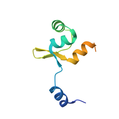
    The recombination directionality factors from Mesorhizobium spp. (RdfS) are involved in regulating the excision and transfer of integrative and conjugative elements. Here, solution small-angle X-ray scattering, and crystallization and preliminary structure solution of RdfS from Mesorhizobium japonicum R7A are presented. RdfS crystallizes in space group P2 1 2 1 2 1 , with evidence of eightfold rotational crystallographic/noncrystallographic symmetry. Initial structure determination by molecular replacement using ab initio models yielded a partial model (three molecules), which was completed after manual inspection revealed unmodelled electron density. The finalized crystal structure of RdfS reveals a head-to-tail polymer forming left-handed superhelices with large solvent channels. Additionally, RdfS has significant disorder in the C-terminal region of the protein, which is supported by the solution scattering data and the crystal structure. The steps taken to finalize structure determination, as well as the scattering and crystallographic characteristics of RdfS, are discussed.


  • Organizational Affiliation
    • School of Molecular Sciences, University of Western Australia, Perth, Western Australia 6009, Australia.

Macromolecule Content 

  • Total Structure Weight: 40.26 kDa 
  • Atom Count: 2,359 
  • Modeled Residue Count: 270 
  • Deposited Residue Count: 364 
  • Unique protein chains: 1

Macromolecules

Find similar proteins by:|  3D Structure
Entity ID: 1
MoleculeChains  Sequence LengthOrganismDetailsImage
Recombination Directionality Factor RdfS
A, B, C, D
91Mesorhizobium japonicum R7AMutation(s): 0 
Gene Names: msi109A8146_15230BAE39_30655EB815_31145
UniProt
Find proteins for Q7AL96 (Rhizobium loti)
Explore Q7AL96 
Go to UniProtKB:  Q7AL96
Entity Groups
Sequence Clusters30% Identity50% Identity70% Identity90% Identity95% Identity100% Identity
UniProt GroupQ7AL96
Sequence Annotations
Expand
Reference Sequence

Experimental Data & Validation

Experimental Data

  • Method: X-RAY DIFFRACTION
  • Resolution: 2.45 Å
  • R-Value Free:  0.246 (Depositor), 0.245 (DCC) 
  • R-Value Work:  0.199 (Depositor), 0.200 (DCC) 
  • R-Value Observed: 0.202 (Depositor) 
Space Group: P 21 21 21
Unit Cell:
Length ( Å )Angle ( ˚ )
a = 35.251α = 90
b = 119.204β = 90
c = 123.397γ = 90
Software Package:
Software NamePurpose
PHENIXrefinement
XDSdata reduction
Aimlessdata scaling
PHASERphasing
MOLREPphasing
PDB_EXTRACTdata extraction

Structure Validation

View Full Validation Report


Entry History 

& Funding Information

Deposition Data


Funding OrganizationLocationGrant Number
Australian Research Council (ARC)AustraliaFT170100235

Revision History  (Full details and data files)

  • Version 1.0: 2022-10-05
    Type: Initial release
  • Version 1.1: 2022-10-12
    Changes: Database references
  • Version 1.2: 2024-04-03
    Changes: Data collection, Refinement description