8DFT | pdb_00008dft

Cryo-EM structure of conjugative pili from Pyrobaculum calidifontis


Experimental Data Snapshot

  • Method: ELECTRON MICROSCOPY
  • Resolution: 4.10 Å
  • Aggregation State: FILAMENT 
  • Reconstruction Method: HELICAL 

wwPDB Validation 3D Report Full Report

Validation slider image for 8DFT

This is version 1.2 of the entry. See complete history

Literature

Archaeal DNA-import apparatus is homologous to bacterial conjugation machinery.

Beltran, L.C.Cvirkaite-Krupovic, V.Miller, J.Wang, F.Kreutzberger, M.A.B.Patkowski, J.B.Costa, T.R.D.Schouten, S.Levental, I.Conticello, V.P.Egelman, E.H.Krupovic, M.

(2023) Nat Commun 14: 666-666

  • DOI: https://doi.org/10.1038/s41467-023-36349-8
  • Primary Citation Related Structures: 
    8DFT, 8DFU, 8EXH

  • PubMed Abstract: 

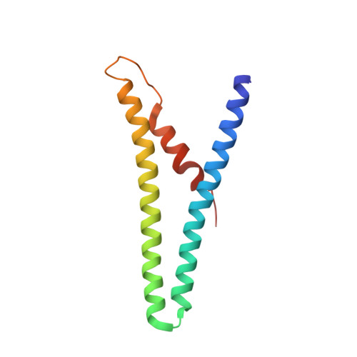
    Conjugation is a major mechanism of horizontal gene transfer promoting the spread of antibiotic resistance among human pathogens. It involves establishing a junction between a donor and a recipient cell via an extracellular appendage known as the mating pilus. In bacteria, the conjugation machinery is encoded by plasmids or transposons and typically mediates the transfer of cognate mobile genetic elements. Much less is known about conjugation in archaea. Here, we determine atomic structures by cryo-electron microscopy of three conjugative pili, two from hyperthermophilic archaea (Aeropyrum pernix and Pyrobaculum calidifontis) and one encoded by the Ti plasmid of the bacterium Agrobacterium tumefaciens, and show that the archaeal pili are homologous to bacterial mating pili. However, the archaeal conjugation machinery, known as Ced, has been 'domesticated', that is, the genes for the conjugation machinery are encoded on the chromosome rather than on mobile genetic elements, and mediates the transfer of cellular DNA.


  • Organizational Affiliation
    • Department of Biochemistry and Molecular Genetics, University of Virginia, Charlottesville, VA, 22903, USA.

Macromolecule Content 

  • Total Structure Weight: 264.91 kDa 
  • Atom Count: 18,740 
  • Modeled Residue Count: 2,240 
  • Deposited Residue Count: 2,240 
  • Unique protein chains: 1

Macromolecules

Find similar proteins by:|  3D Structure
Entity ID: 1
MoleculeChains  Sequence LengthOrganismDetailsImage
Pilin protein
A, B, C, D, E
A, B, C, D, E, F, G, H, I, J, K, L, M, N, O, P, Q, R, S, T
112Pyrobaculum calidifontisMutation(s): 0 
Membrane Entity: Yes 
UniProt
Find proteins for A3MU74 (Pyrobaculum calidifontis (strain DSM 21063 / JCM 11548 / VA1))
Explore A3MU74 
Go to UniProtKB:  A3MU74
Entity Groups
Sequence Clusters30% Identity50% Identity70% Identity90% Identity95% Identity100% Identity
UniProt GroupA3MU74
Sequence Annotations
Expand
Reference Sequence

Small Molecules

Ligands 1 Unique
IDChains Name / Formula / InChI Key2D Diagram3D Interactions
TT0
(Subject of Investigation/LOI)

Query on TT0


Download:Ideal Coordinates CCD File
AA [auth G]
BA [auth H]
CA [auth I]
DA [auth J]
EA [auth K]
AA [auth G],
BA [auth H],
CA [auth I],
DA [auth J],
EA [auth K],
FA [auth L],
GA [auth M],
HA [auth N],
IA [auth O],
JA [auth P],
KA [auth Q],
LA [auth R],
MA [auth S],
NA [auth T],
U [auth A],
V [auth B],
W [auth C],
X [auth D],
Y [auth E],
Z [auth F]
[(2~{S},7~{S},11~{S},15~{S},19~{R},22~{R},26~{S},30~{R},34~{R},38~{S},43~{S},47~{S},51~{S},55~{R},58~{R},62~{S},66~{R},70~{R})-38-(hydroxymethyl)-7,11,15,19,22,26,30,34,43,47,51,55,58,62,66,70-hexadecamethyl-1,4,37,40-tetraoxacyclodoheptacont-2-yl]methanol
C86 H172 O6
VMHUDYKDOMRJOK-PFTGJOHNSA-N

Experimental Data & Validation

Experimental Data

  • Method: ELECTRON MICROSCOPY
  • Resolution: 4.10 Å
  • Aggregation State: FILAMENT 
  • Reconstruction Method: HELICAL 
EM Software:
TaskSoftware PackageVersion
RECONSTRUCTIONcryoSPARC3.2

Structure Validation

View Full Validation Report


Entry History 

& Funding Information

Deposition Data


Funding OrganizationLocationGrant Number
National Institutes of Health/National Institute of General Medical Sciences (NIH/NIGMS)United StatesGM122510

Revision History  (Full details and data files)

  • Version 1.0: 2023-03-22
    Type: Initial release
  • Version 1.1: 2024-06-12
    Changes: Data collection
  • Version 1.2: 2024-09-25
    Changes: Data collection, Experimental preparation