8DDR

cryo-EM structure of TRPM3 ion channel in the absence of PIP2


Experimental Data Snapshot

  • Method: ELECTRON MICROSCOPY
  • Resolution: 3.20 Å
  • Aggregation State: PARTICLE 
  • Reconstruction Method: SINGLE PARTICLE 

wwPDB Validation   3D Report Full Report


This is version 1.2 of the entry. See complete history


Literature

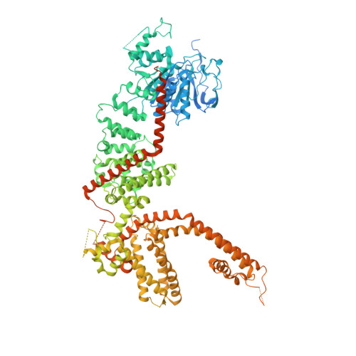
Structural and functional analyses of a GPCR-inhibited ion channel TRPM3.

Zhao, C.MacKinnon, R.

(2023) Neuron 111: 81

  • DOI: https://doi.org/10.1016/j.neuron.2022.10.002
  • Primary Citation of Related Structures:  
    8DDQ, 8DDR, 8DDS, 8DDT, 8DDU, 8DDV, 8DDW, 8DDX, 8ED7, 8ED8, 8ED9

  • PubMed Abstract: 

    G-protein coupled receptors (GPCRs) govern the physiological response to stimuli by modulating the activity of downstream effectors, including ion channels. TRPM3 is an ion channel inhibited by GPCRs through direct interaction with G protein (Gβγ) released upon their activation. This GPCR-TRPM3 signaling pathway contributes to the analgesic effect of morphine. Here, we characterized Gβγ inhibition of TRPM3 using electrophysiology and single particle cryo-electron microscopy (cryo-EM). From electrophysiology, we obtained a half inhibition constant (IC50) of ∼240 nM. Using cryo-EM, we determined structures of mouse TRPM3 expressed in human cells with and without Gβγ and with and without PIP 2 , a lipid required for TRPM3 activity, at resolutions of 2.7-4.7 Å. Gβγ-TRPM3 interfaces vary depending on PIP 2 occupancy; however, in all cases, Gβγ appears loosely attached to TRPM3. The IC50 in electrophysiology experiments raises the possibility that additional unknown factors may stabilize the TRPM3-Gβγ complex.


  • Organizational Affiliation

    Laboratory of Molecular Neurobiology and Biophysics, Howard Hughes Medical Institute, the Rockefeller University, New York, NY 10065, United States.


Macromolecules
Find similar proteins by:  (by identity cutoff)  |  3D Structure
Entity ID: 1
MoleculeChains Sequence LengthOrganismDetailsImage
Transient receptor potential cation channel, subfamily M, member 3A,
C [auth B],
E [auth C],
G [auth D]
1,343Mus musculusMutation(s): 0 
Gene Names: Trpm3
Membrane Entity: Yes 
UniProt & NIH Common Fund Data Resources
Find proteins for Q5F4S7 (Mus musculus)
Explore Q5F4S7 
Go to UniProtKB:  Q5F4S7
IMPC:  MGI:2443101
Entity Groups  
Sequence Clusters30% Identity50% Identity70% Identity90% Identity95% Identity100% Identity
UniProt GroupQ5F4S7
Sequence Annotations
Expand
  • Reference Sequence

Find similar proteins by:  Sequence   |   3D Structure  

Entity ID: 2
MoleculeChains Sequence LengthOrganismDetailsImage
Unidentified segment at the N-terminus of TRPM3B [auth E],
D [auth F],
F [auth G],
H
17Mus musculusMutation(s): 0 
Sequence Annotations
Expand
  • Reference Sequence
Small Molecules
Ligands 3 Unique
IDChains Name / Formula / InChI Key2D Diagram3D Interactions
3PH
Query on 3PH

Download Ideal Coordinates CCD File 
I [auth A],
N [auth B],
P [auth C],
R [auth D]
1,2-DIACYL-GLYCEROL-3-SN-PHOSPHATE
C39 H77 O8 P
YFWHNAWEOZTIPI-DIPNUNPCSA-N
9Z9
Query on 9Z9

Download Ideal Coordinates CCD File 
J [auth A],
M [auth B],
O [auth C],
Q [auth D]
(3beta,14beta,17beta,25R)-3-[4-methoxy-3-(methoxymethyl)butoxy]spirost-5-en
C34 H56 O5
CEEBZAXXSRFQIC-GZSGZGDASA-N
NA
Query on NA

Download Ideal Coordinates CCD File 
K [auth A],
L [auth A]
SODIUM ION
Na
FKNQFGJONOIPTF-UHFFFAOYSA-N
Experimental Data & Validation

Experimental Data

  • Method: ELECTRON MICROSCOPY
  • Resolution: 3.20 Å
  • Aggregation State: PARTICLE 
  • Reconstruction Method: SINGLE PARTICLE 

Structure Validation

View Full Validation Report


Entry History & Funding Information

Deposition Data


Funding OrganizationLocationGrant Number
Howard Hughes Medical Institute (HHMI)United States--

Revision History  (Full details and data files)

  • Version 1.0: 2022-11-02
    Type: Initial release
  • Version 1.1: 2022-11-09
    Changes: Database references
  • Version 1.2: 2023-01-18
    Changes: Database references