8D6V

Structure of the Mycobacterium tuberculosis 20S proteasome bound to the C-terminal GQYL motif of the ATP-bound Mpa ATPase


Experimental Data Snapshot

  • Method: ELECTRON MICROSCOPY
  • Resolution: 3.20 Å
  • Aggregation State: PARTICLE 
  • Reconstruction Method: SINGLE PARTICLE 

wwPDB Validation   3D Report Full Report


This is version 1.2 of the entry. See complete history


Literature

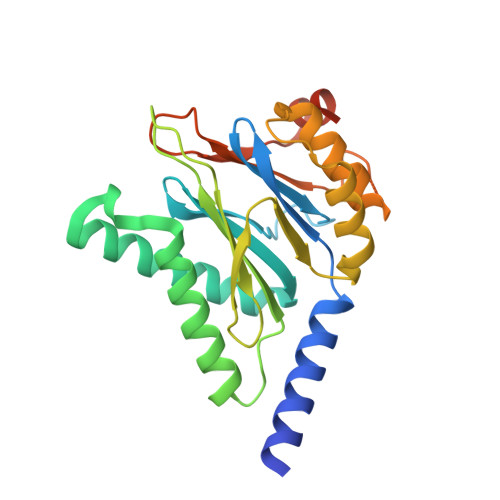
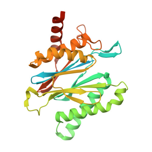
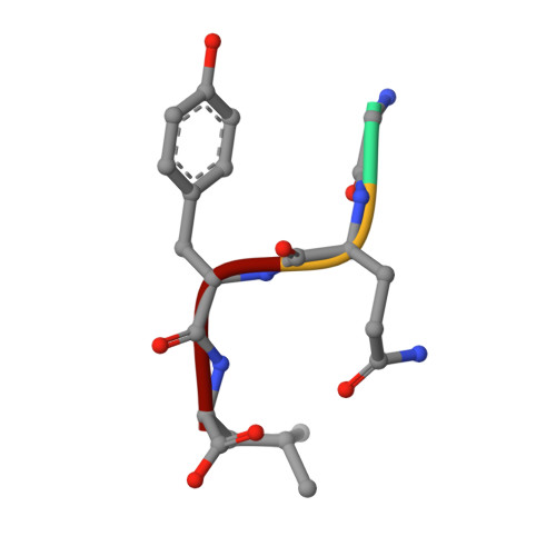
The beta-Grasp Domain of Proteasomal ATPase Mpa Makes Critical Contacts with the Mycobacterium tuberculosis 20S Core Particle to Facilitate Degradation.

Xiao, X.Feng, X.Yoo, J.H.Kovach, A.Darwin, K.H.Li, H.

(2022) mSphere 7: e0027422-e0027422

  • DOI: https://doi.org/10.1128/msphere.00274-22
  • Primary Citation of Related Structures:  
    8D6V, 8D6W, 8D6X, 8D6Y

  • PubMed Abstract: 

    Mycobacterium tuberculosis possesses a Pup-proteasome system analogous to the eukaryotic ubiquitin-proteasome pathway. We have previously shown that the hexameric mycobacterial proteasome ATPase (Mpa) recruits pupylated protein substrates via interactions between amino-terminal coiled-coils in Mpa monomers and the degradation tag Pup. However, it is unclear how Mpa rings interact with a proteasome due to the presence of a carboxyl-terminal β-grasp domain unique to Mpa homologues that makes the interaction highly unstable. Here, we describe newly identified critical interactions between Mpa and 20S core proteasomes. Interestingly, the Mpa C-terminal GQYL motif binds the 20S core particle activation pocket differently than the same motif of the ATP-independent proteasome accessory factor PafE. We further found that the β-hairpin of the Mpa β-grasp domain interacts variably with the H0 helix on top of the 20S core particle via a series of ionic and hydrogen-bond interactions. Individually mutating several involved residues reduced Mpa-mediated protein degradation both in vitro and in vivo . IMPORTANCE The Pup-proteasome system in Mycobacterium tuberculosis is critical for this species to cause lethal infections in mice. Investigating the molecular mechanism of how the Mpa ATPase recruits and unfolds pupylated substrates to the 20S proteasomal core particle for degradation will be essential to fully understand how degradation is regulated, and the structural information we report may be useful for the development of new tuberculosis chemotherapies.


  • Organizational Affiliation

    Department of Structural Biology, Van Andel Institutegrid.251017.0, Grand Rapids, Michigan, USA.


Macromolecules
Find similar proteins by:  (by identity cutoff)  |  3D Structure
Entity ID: 1
MoleculeChains Sequence LengthOrganismDetailsImage
Proteasome subunit alpha248Mycobacterium tuberculosisMutation(s): 0 
Gene Names: prcAMRA_2124
EC: 3.4.25.1
UniProt
Find proteins for P9WHU1 (Mycobacterium tuberculosis (strain ATCC 25618 / H37Rv))
Explore P9WHU1 
Go to UniProtKB:  P9WHU1
Entity Groups  
Sequence Clusters30% Identity50% Identity70% Identity90% Identity95% Identity100% Identity
UniProt GroupP9WHU1
Sequence Annotations
Expand
  • Reference Sequence
Find similar proteins by:  (by identity cutoff)  |  3D Structure
Entity ID: 2
MoleculeChains Sequence LengthOrganismDetailsImage
Proteasome subunit beta291Mycobacterium tuberculosisMutation(s): 0 
Gene Names: 
EC: 3.4.25.1
UniProt
Find proteins for P9WHT9 (Mycobacterium tuberculosis (strain ATCC 25618 / H37Rv))
Explore P9WHT9 
Go to UniProtKB:  P9WHT9
Entity Groups  
Sequence Clusters30% Identity50% Identity70% Identity90% Identity95% Identity100% Identity
UniProt GroupP9WHT9
Sequence Annotations
Expand
  • Reference Sequence

Find similar proteins by:  Sequence   |   3D Structure  

Entity ID: 3
MoleculeChains Sequence LengthOrganismDetailsImage
Proteasome-associated ATPase4Mycobacterium tuberculosisMutation(s): 0 
Gene Names: mpaBCG_2132c
UniProt
Find proteins for P9WQN5 (Mycobacterium tuberculosis (strain ATCC 25618 / H37Rv))
Explore P9WQN5 
Go to UniProtKB:  P9WQN5
Entity Groups  
UniProt GroupP9WQN5
Sequence Annotations
Expand
  • Reference Sequence
Experimental Data & Validation

Experimental Data

  • Method: ELECTRON MICROSCOPY
  • Resolution: 3.20 Å
  • Aggregation State: PARTICLE 
  • Reconstruction Method: SINGLE PARTICLE 

Structure Validation

View Full Validation Report


Entry History & Funding Information

Deposition Data


Funding OrganizationLocationGrant Number
National Institutes of Health/National Library of Medicine (NIH/NLM)United StatesR01

Revision History  (Full details and data files)

  • Version 1.0: 2022-08-03
    Type: Initial release
  • Version 1.1: 2022-09-07
    Changes: Database references
  • Version 1.2: 2022-11-09
    Changes: Database references