Synthesis of C 7 /C 8 -cyclitols and C 7 N-aminocyclitols from maltose and X-ray crystal structure of Streptomyces coelicolor GlgEI V279S in a complex with an amylostatin GXG-like derivative.
Thanvi, R., Jayasinghe, T.D., Kapil, S., Obadawo, B.S., Ronning, D.R., Sucheck, S.J.(2022) Front Chem 10: 950433-950433
- PubMed: 36157042 Search on PubMedSearch on PubMed Central
- DOI: https://doi.org/10.3389/fchem.2022.950433
- Primary Citation Related Structures:
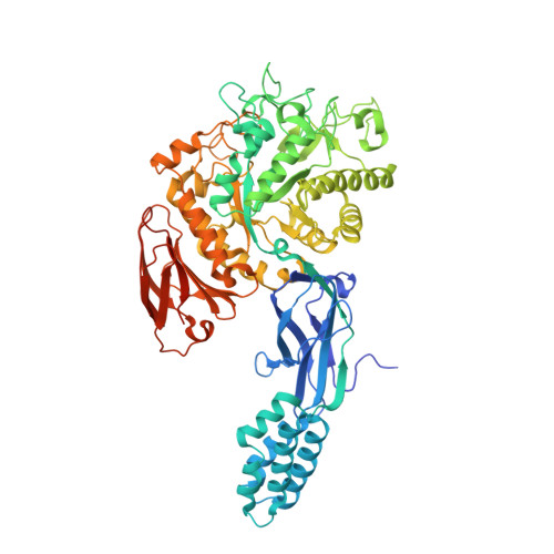
8D6K - PubMed Abstract:
C 7 /C 8 -cyclitols and C 7 N-aminocyclitols find applications in the pharmaceutical sector as α-glucosidase inhibitors and in the agricultural sector as fungicides and insecticides. In this study, we identified C 7 /C 8 -cyclitols and C 7 N-aminocyclitols as potential inhibitors of Streptomyces coelicolor ( Sco ) GlgEI-V279S based on the docking scores. The protein and the ligand (targets 11 , 12 , and 13 ) were prepared, the states were generated at pH 7.0 ± 2.0, and the ligands were docked into the active sites of the receptor via Glide™. The synthetic route to these targets was similar to our previously reported route used to obtain 4-⍺-glucoside of valienamine ( AGV ), except the protecting group for target 12 was a p -bromobenzyl (PBB) ether to preserve the alkene upon deprotection. While compounds 11 - 13 did not inhibit Sco GlgEI-V279S at the concentrations evaluated, an X-ray crystal structure of the Sco GlgE1-V279S/ 13 complex was solved to a resolution of 2.73 Å. This structure allowed assessment differences and commonality with our previously reported inhibitors and was useful for identifying enzyme-compound interactions that may be important for future inhibitor development. The Asp 394 nucleophile formed a bidentate hydrogen bond interaction with the exocyclic oxygen atoms (C(3)-OH and C(7)-OH) similar to the observed interactions with the Sco GlgEI-V279S in a complex with AGV (PDB:7MGY). In addition, the data suggest replacing the cyclohexyl group with more isosteric and hydrogen bond-donating groups to increase binding interactions in the + 1 binding site.
- Department of Chemistry and Biochemistry, The University of Toledo, Toledo, OH, United States.
Organizational Affiliation: