


Crystal structure of WhiB6 and SigA4-betaTip complex
Wan, T., Zhang, L.M.To be published.
Experimental Data Snapshot
Starting Model: experimental
View more details
wwPDB Validation 3D Report Full Report

Entity ID: 1 | |||||
|---|---|---|---|---|---|
| Molecule | Chains | Sequence Length | Organism | Details | Image |
| Probable transcriptional regulator WhiB6 | 116 | Mycobacterium tuberculosis | Mutation(s): 0 Gene Names: whiB6, Rv3862c | 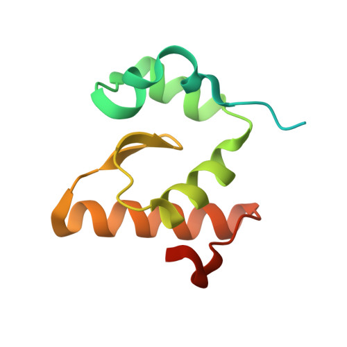 | |
UniProt | |||||
Entity Groups | |||||
| Sequence Clusters | 30% Identity50% Identity70% Identity90% Identity95% Identity100% Identity | ||||
| UniProt Group | P9WF37 | ||||
Sequence AnnotationsExpand | |||||
Reference Sequence | |||||
Entity ID: 2 | |||||
|---|---|---|---|---|---|
| Molecule | Chains | Sequence Length | Organism | Details | Image |
| RNA polymerase sigma factor SigA,DNA-directed RNA polymerase subunit beta | 112 | Mycobacterium tuberculosis H37Rv, Mycobacterium tuberculosis This entity is chimeric | Mutation(s): 0 Gene Names: sigA, mysA, rpoD, rpoV, Rv2703, MTCY05A6.24, rpoB, Rv0667, MTCI376.08c EC: 2.7.7.6 |  | |
UniProt | |||||
Entity Groups | |||||
| Sequence Clusters | 30% Identity50% Identity70% Identity90% Identity95% Identity100% Identity | ||||
| UniProt Groups | P9WGY9P9WGI1 | ||||
Sequence AnnotationsExpand | |||||
Reference Sequence | |||||
| Ligands 1 Unique | |||||
|---|---|---|---|---|---|
| ID | Chains | Name / Formula / InChI Key | 2D Diagram | 3D Interactions | |
| SF4 (Subject of Investigation/LOI) Download:Ideal Coordinates CCD File | E [auth A], F [auth C] | IRON/SULFUR CLUSTER Fe4 S4 LJBDFODJNLIPKO-UHFFFAOYSA-N |  | ||
| Length ( Å ) | Angle ( ˚ ) |
|---|---|
| a = 62.983 | α = 90 |
| b = 66.54 | β = 90 |
| c = 158.805 | γ = 90 |
| Software Name | Purpose |
|---|---|
| PHENIX | refinement |
| XDS | data reduction |
| SCALA | data scaling |
| PHASER | phasing |
| Funding Organization | Location | Grant Number |
|---|---|---|
| National Institutes of Health/National Institute of General Medical Sciences (NIH/NIGMS) | United States | R35 GM138157-01 |
| National Science Foundation (NSF, United States) | United States | CLP 1846908 |


