A Noble Metal Substitution Leads to B 12 Cofactor Mimicry by a Rhodibalamin.
Ruetz, M., Mascarenhas, R., Widner, F., Kieninger, C., Koutmos, M., Krautler, B., Banerjee, R.(2024) Biochemistry 63: 1955-1962
- PubMed: 39012171 Search on PubMedSearch on PubMed Central
- DOI: https://doi.org/10.1021/acs.biochem.4c00216
- Primary Citation Related Structures:
8D32 - PubMed Abstract:
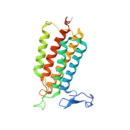
In mammals, cobalamin is an essential cofactor that is delivered by a multitude of chaperones in an elaborate trafficking pathway to two client enzymes, methionine synthase and methylmalonyl-CoA mutase (MMUT). Rhodibalamins, the rhodium analogs of cobalamins, have been described as antimetabolites due to their ability to inhibit bacterial growth. In this study, we have examined the reactivity of adenosylrhodibalamin (AdoRhbl) with two key human chaperones, MMACHC (also known as CblC) and adenosyltransferase (MMAB, also known as ATR), and with the human and Mycobacterium tuberculosis MMUT. We demonstrate that while AdoRhbl binds tightly to all four proteins, the Rh-carbon bond is resistant to homolytic (on MMAB and MMUT) as well as heterolytic (on MMACHC) rupture. On the other hand, MMAB catalyzes Rh-carbon bond formation, converting rhodi(I)balamin in the presence of ATP to AdoRhbl. We report the first crystal structure of a rhodibalamin (AdoRhbl) bound to a B 12 protein, i.e., MMAB, in the presence of triphosphate, which shows a weakened but intact Rh-carbon bond. The structure provides insights into how MMAB cleaves the corresponding Co-carbon bond in a sacrificial homolytic reaction that purportedly functions as a cofactor sequestration strategy. Collectively, the study demonstrates that while the noble metal substitution of cobalt by rhodium sets up structural mimicry, it compromises chemistry, which could be exploited for targeting human and bacterial B 12 chaperones and enzymes.