


Crystal structure of the human COPB2 WD-domains
Zeng, H., Dong, A., Hutchinson, A., Seitova, A., Loppnau, P., Arrowsmith, C.H., Edwards, A.M., Halabelian, L., Structural Genomics Consortium (SGC)To be published.
Experimental Data Snapshot
Starting Model: experimental
View more details
wwPDB Validation 3D Report Full Report

Entity ID: 1 | |||||
|---|---|---|---|---|---|
| Molecule | Chains | Sequence Length | Organism | Details | Image |
| Coatomer subunit beta' | 615 | Homo sapiens | Mutation(s): 0 Gene Names: COPB2 | 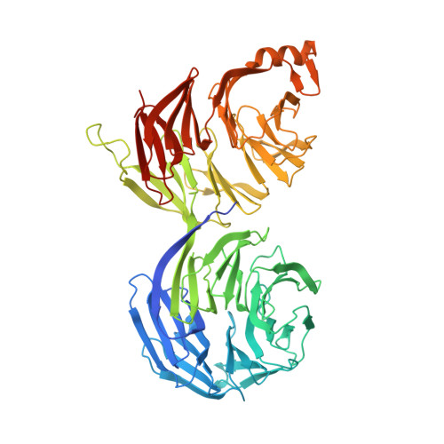 | |
UniProt & NIH Common Fund Data Resources | |||||
PHAROS: P35606 GTEx: ENSG00000184432 | |||||
Entity Groups | |||||
| Sequence Clusters | 30% Identity50% Identity70% Identity90% Identity95% Identity100% Identity | ||||
| UniProt Group | P35606 | ||||
Sequence AnnotationsExpand | |||||
Reference Sequence | |||||
| Ligands 1 Unique | |||||
|---|---|---|---|---|---|
| ID | Chains | Name / Formula / InChI Key | 2D Diagram | 3D Interactions | |
| EDO Download:Ideal Coordinates CCD File | C [auth A] | 1,2-ETHANEDIOL C2 H6 O2 LYCAIKOWRPUZTN-UHFFFAOYSA-N |  | ||
| Length ( Å ) | Angle ( ˚ ) |
|---|---|
| a = 67.074 | α = 90 |
| b = 134.062 | β = 90 |
| c = 144.27 | γ = 90 |
| Software Name | Purpose |
|---|---|
| BUSTER | refinement |
| SCALEPACK | data scaling |
| PDB_EXTRACT | data extraction |
| HKL-3000 | data reduction |
| PHASER | phasing |
| Funding Organization | Location | Grant Number |
|---|---|---|
| Other private | -- |


