8CUY

ACP1-KS-AT domains of mycobacterial Pks13


Experimental Data Snapshot

  • Method: ELECTRON MICROSCOPY
  • Resolution: 2.40 Å
  • Aggregation State: PARTICLE 
  • Reconstruction Method: SINGLE PARTICLE 

wwPDB Validation   3D Report Full Report


This is version 1.2 of the entry. See complete history


Literature

Structure and dynamics of the essential endogenous mycobacterial polyketide synthase Pks13.

Kim, S.K.Dickinson, M.S.Finer-Moore, J.Guan, Z.Kaake, R.M.Echeverria, I.Chen, J.Pulido, E.H.Sali, A.Krogan, N.J.Rosenberg, O.S.Stroud, R.M.

(2023) Nat Struct Mol Biol 30: 296-308

  • DOI: https://doi.org/10.1038/s41594-022-00918-0
  • Primary Citation of Related Structures:  
    7UK4, 8CUY, 8CUZ, 8CV0, 8CV1

  • PubMed Abstract: 

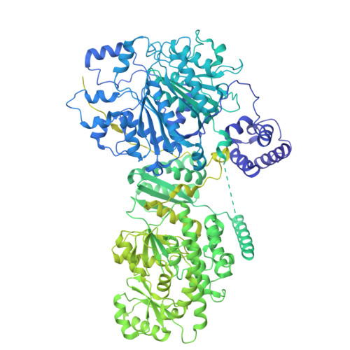
    The mycolic acid layer of the Mycobacterium tuberculosis cell wall is essential for viability and virulence, and the enzymes responsible for its synthesis are targets for antimycobacterial drug development. Polyketide synthase 13 (Pks13) is a module encoding several enzymatic and transport functions that carries out the condensation of two different long-chain fatty acids to produce mycolic acids. We determined structures by cryogenic-electron microscopy of dimeric multi-enzyme Pks13 purified from mycobacteria under normal growth conditions, captured with native substrates. Structures define the ketosynthase (KS), linker and acyl transferase (AT) domains at 1.8 Å resolution and two alternative locations of the N-terminal acyl carrier protein. These structures suggest intermediate states on the pathway for substrate delivery to the KS domain. Other domains, visible at lower resolution, are flexible relative to the KS-AT core. The chemical structures of three bound endogenous long-chain fatty acid substrates were determined by electrospray ionization mass spectrometry.


  • Organizational Affiliation

    Department of Biochemistry and Biophysics, University of California San Francisco, San Francisco, CA, USA.


Macromolecules
Find similar proteins by:  (by identity cutoff)  |  3D Structure
Entity ID: 1
MoleculeChains Sequence LengthOrganismDetailsImage
Polyketide synthase PKS13
A, B
1,816Mycolicibacterium smegmatis MC2 155Mutation(s): 0 
EC: 2.3.1.94
UniProt
Find proteins for I7FMV0 (Mycolicibacterium smegmatis (strain ATCC 700084 / mc(2)155))
Explore I7FMV0 
Go to UniProtKB:  I7FMV0
Entity Groups  
Sequence Clusters30% Identity50% Identity70% Identity90% Identity95% Identity100% Identity
UniProt GroupI7FMV0
Sequence Annotations
Expand
  • Reference Sequence
Experimental Data & Validation

Experimental Data

  • Method: ELECTRON MICROSCOPY
  • Resolution: 2.40 Å
  • Aggregation State: PARTICLE 
  • Reconstruction Method: SINGLE PARTICLE 
EM Software:
TaskSoftware PackageVersion
MODEL REFINEMENTISOLDE
MODEL REFINEMENTPHENIX1.19.2
RECONSTRUCTIONRELION3.0

Structure Validation

View Full Validation Report


Entry History & Funding Information

Deposition Data


Funding OrganizationLocationGrant Number
National Institutes of Health/National Institute Of Allergy and Infectious Diseases (NIH/NIAID)United StatesP01 AI095208
National Institutes of Health/National Institute Of Allergy and Infectious Diseases (NIH/NIAID)United StatesR01 AI128214
National Institutes of Health/National Institute of General Medical Sciences (NIH/NIGMS)United StatesR01 GM24485

Revision History  (Full details and data files)

  • Version 1.0: 2023-02-15
    Type: Initial release
  • Version 1.1: 2023-03-01
    Changes: Database references
  • Version 1.2: 2023-03-29
    Changes: Database references