8CTK | pdb_00008ctk

Cryo-EM structure of SARS-CoV-2 M protein in a lipid nanodisc


Experimental Data Snapshot

  • Method: ELECTRON MICROSCOPY
  • Resolution: 3.52 Å
  • Aggregation State: PARTICLE 
  • Reconstruction Method: SINGLE PARTICLE 

wwPDB Validation 3D Report Full Report

Validation slider image for 8CTK

This is version 1.2 of the entry. See complete history

Literature

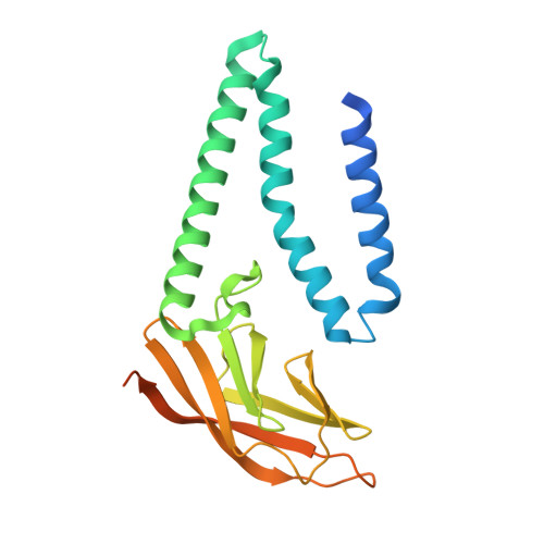
Structure of SARS-CoV-2 M protein in lipid nanodiscs.

Dolan, K.A.Dutta, M.Kern, D.M.Kotecha, A.Voth, G.A.Brohawn, S.G.

(2022) Elife 11

  • DOI: https://doi.org/10.7554/eLife.81702
  • Primary Citation Related Structures: 
    8CTK

  • PubMed Abstract: 

    SARS-CoV-2 encodes four structural proteins incorporated into virions, spike (S), envelope (E), nucleocapsid (N), and membrane (M). M plays an essential role in viral assembly by organizing other structural proteins through physical interactions and directing them to sites of viral budding. As the most abundant protein in the viral envelope and a target of patient antibodies, M is a compelling target for vaccines and therapeutics. Still, the structure of M and molecular basis for its role in virion formation are unknown. Here, we present the cryo-EM structure of SARS-CoV-2 M in lipid nanodiscs to 3.5 Å resolution. M forms a 50 kDa homodimer that is structurally related to the SARS-CoV-2 ORF3a viroporin, suggesting a shared ancestral origin. Structural comparisons reveal how intersubunit gaps create a small, enclosed pocket in M and large open cavity in ORF3a, consistent with a structural role and ion channel activity, respectively. M displays a strikingly electropositive cytosolic surface that may be important for interactions with N, S, and viral RNA. Molecular dynamics simulations show a high degree of structural rigidity in a simple lipid bilayer and support a role for M homodimers in scaffolding viral assembly. Together, these results provide insight into roles for M in coronavirus assembly and structure.


  • Organizational Affiliation
    • Biophysics Graduate Group, University of California, Berkeley, Berkeley, United States.

Macromolecule Content 

  • Total Structure Weight: 52.38 kDa 
  • Atom Count: 3,042 
  • Modeled Residue Count: 376 
  • Deposited Residue Count: 462 
  • Unique protein chains: 1

Macromolecules

Find similar proteins by:|  3D Structure
Entity ID: 1
MoleculeChains  Sequence LengthOrganismDetailsImage
Membrane protein
A, B
231Severe acute respiratory syndrome coronavirus 2Mutation(s): 0 
Membrane Entity: Yes 
UniProt
Find proteins for P0DTC5 (Severe acute respiratory syndrome coronavirus 2)
Explore P0DTC5 
Go to UniProtKB:  P0DTC5
Entity Groups
Sequence Clusters30% Identity50% Identity70% Identity90% Identity95% Identity100% Identity
UniProt GroupP0DTC5
Sequence Annotations
Expand
Reference Sequence

Experimental Data & Validation

Experimental Data

  • Method: ELECTRON MICROSCOPY
  • Resolution: 3.52 Å
  • Aggregation State: PARTICLE 
  • Reconstruction Method: SINGLE PARTICLE 
EM Software:
TaskSoftware PackageVersion
RECONSTRUCTIONcryoSPARC3.1

Structure Validation

View Full Validation Report


Entry History 

& Funding Information

Deposition Data


Funding OrganizationLocationGrant Number
National Institutes of Health/National Institute of General Medical Sciences (NIH/NIGMS)United StatesGM123496

Revision History  (Full details and data files)

  • Version 1.0: 2022-06-22
    Type: Initial release
  • Version 1.1: 2022-11-02
    Changes: Database references
  • Version 1.2: 2024-06-12
    Changes: Data collection