Structural basis of NINJ1-mediated plasma membrane rupture in cell death.
Degen, M., Santos, J.C., Pluhackova, K., Cebrero, G., Ramos, S., Jankevicius, G., Hartenian, E., Guillerm, U., Mari, S.A., Kohl, B., Muller, D.J., Schanda, P., Maier, T., Perez, C., Sieben, C., Broz, P., Hiller, S.(2023) Nature 618: 1065-1071
- PubMed: 37198476 Search on PubMedSearch on PubMed Central
- DOI: https://doi.org/10.1038/s41586-023-05991-z
- Primary Citation Related Structures:
8CQR - PubMed Abstract:
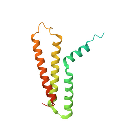
Eukaryotic cells can undergo different forms of programmed cell death, many of which culminate in plasma membrane rupture as the defining terminal event 1-7 . Plasma membrane rupture was long thought to be driven by osmotic pressure, but it has recently been shown to be in many cases an active process, mediated by the protein ninjurin-1 8 (NINJ1). Here we resolve the structure of NINJ1 and the mechanism by which it ruptures membranes. Super-resolution microscopy reveals that NINJ1 clusters into structurally diverse assemblies in the membranes of dying cells, in particular large, filamentous assemblies with branched morphology. A cryo-electron microscopy structure of NINJ1 filaments shows a tightly packed fence-like array of transmembrane α-helices. Filament directionality and stability is defined by two amphipathic α-helices that interlink adjacent filament subunits. The NINJ1 filament features a hydrophilic side and a hydrophobic side, and molecular dynamics simulations show that it can stably cap membrane edges. The function of the resulting supramolecular arrangement was validated by site-directed mutagenesis. Our data thus suggest that, during lytic cell death, the extracellular α-helices of NINJ1 insert into the plasma membrane to polymerize NINJ1 monomers into amphipathic filaments that rupture the plasma membrane. The membrane protein NINJ1 is therefore an interactive component of the eukaryotic cell membrane that functions as an in-built breaking point in response to activation of cell death.
- Biozentrum, University of Basel, Basel, Switzerland.
Organizational Affiliation: