8CP2

Structure of Aspartate-N-hydroxylase (FzmM)from Streptomyces sp. V2: complex with NADPH and L-aspartate


Experimental Data Snapshot

  • Method: X-RAY DIFFRACTION
  • Resolution: 2.10 Å
  • R-Value Free: 0.202 
  • R-Value Work: 0.179 
  • R-Value Observed: 0.180 

wwPDB Validation   3D Report Full Report


Ligand Structure Quality Assessment 


This is version 1.1 of the entry. See complete history


Literature

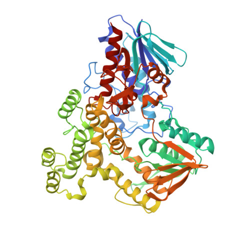
A biosynthetic aspartate N-hydroxylase performs successive oxidations by holding intermediates at a site away from the catalytic center.

Rotilio, L.Boverio, A.Nguyen, Q.T.Mannucci, B.Fraaije, M.W.Mattevi, A.

(2023) J Biol Chem 299: 104904-104904

  • DOI: https://doi.org/10.1016/j.jbc.2023.104904
  • Primary Citation of Related Structures:  
    8CP2, 8CP5

  • PubMed Abstract: 

    Nitrosuccinate is a biosynthetic building block in many microbial pathways. The metabolite is produced by dedicated L-aspartate hydroxylases that use NADPH and molecular oxygen as co-substrates. Here, we investigate the mechanism underlying the unusual ability of these enzymes to perform successive rounds of oxidative modifications. The crystal structure of Streptomyces sp. V2 L-aspartate N-hydroxylase outlines a characteristic helical domain wedged between two dinucleotide-binding domains. Together with NADPH and FAD, a cluster of conserved arginine residues forms the catalytic core at the domain interface. Aspartate is found to bind in an entry chamber that is close to but not in direct contact with the flavin. It is recognized by an extensive H-bond network that explains the enzyme's strict substrate-selectivity. A mutant designed to create steric and electrostatic hindrance to substrate binding disables hydroxylation without perturbing the NADPH oxidase side-activity. Critically, the distance between the FAD and the substrate is far too long to afford N-hydroxylation by the C4a-hydroperoxyflavin intermediate whose formation is confirmed by our work. We conclude that the enzyme functions through a catch-and-release mechanism. L-aspartate slides into the catalytic center only when the hydroxylating apparatus is formed. It is then re-captured by the entry chamber where it waits for the next round of hydroxylation. By iterating these steps, the enzyme minimizes the leakage of incompletely oxygenated products and ensures that the reaction carries on until nitrosuccinate is formed. This unstable product can then be engaged by a successive biosynthetic enzyme or undergoes spontaneous decarboxylation to produce 3-nitropropionate, a mycotoxin.


  • Organizational Affiliation

    Department of Biology and Biotechnology, University of Pavia, Pavia, Italy.


Macromolecules
Find similar proteins by:  (by identity cutoff)  |  3D Structure
Entity ID: 1
MoleculeChains Sequence LengthOrganismDetailsImage
FAD-binding protein
A, B
601Streptomyces sp. V2Mutation(s): 0 
Gene Names: DF268_28005
UniProt
Find proteins for A0A2V1NMV1 (Streptomyces sp. V2)
Explore A0A2V1NMV1 
Go to UniProtKB:  A0A2V1NMV1
Entity Groups  
Sequence Clusters30% Identity50% Identity70% Identity90% Identity95% Identity100% Identity
UniProt GroupA0A2V1NMV1
Sequence Annotations
Expand
  • Reference Sequence
Small Molecules
Ligands 6 Unique
IDChains Name / Formula / InChI Key2D Diagram3D Interactions
FAD (Subject of Investigation/LOI)
Query on FAD

Download Ideal Coordinates CCD File 
C [auth A],
L [auth B]
FLAVIN-ADENINE DINUCLEOTIDE
C27 H33 N9 O15 P2
VWWQXMAJTJZDQX-UYBVJOGSSA-N
NAP (Subject of Investigation/LOI)
Query on NAP

Download Ideal Coordinates CCD File 
D [auth A],
M [auth B]
NADP NICOTINAMIDE-ADENINE-DINUCLEOTIDE PHOSPHATE
C21 H28 N7 O17 P3
XJLXINKUBYWONI-NNYOXOHSSA-N
ASP (Subject of Investigation/LOI)
Query on ASP

Download Ideal Coordinates CCD File 
E [auth A],
F [auth A],
N [auth B]
ASPARTIC ACID
C4 H7 N O4
CKLJMWTZIZZHCS-REOHCLBHSA-N
3NP
Query on 3NP

Download Ideal Coordinates CCD File 
G [auth A]3-NITROPROPANOIC ACID
C3 H5 N O4
WBLZUCOIBUDNBV-UHFFFAOYSA-N
PEG
Query on PEG

Download Ideal Coordinates CCD File 
H [auth A]
I [auth A]
J [auth A]
O [auth B]
P [auth B]
H [auth A],
I [auth A],
J [auth A],
O [auth B],
P [auth B],
Q [auth B]
DI(HYDROXYETHYL)ETHER
C4 H10 O3
MTHSVFCYNBDYFN-UHFFFAOYSA-N
GOL
Query on GOL

Download Ideal Coordinates CCD File 
K [auth A]GLYCEROL
C3 H8 O3
PEDCQBHIVMGVHV-UHFFFAOYSA-N
Experimental Data & Validation

Experimental Data

  • Method: X-RAY DIFFRACTION
  • Resolution: 2.10 Å
  • R-Value Free: 0.202 
  • R-Value Work: 0.179 
  • R-Value Observed: 0.180 
  • Space Group: H 3
Unit Cell:
Length ( Å )Angle ( ˚ )
a = 244.591α = 90
b = 244.591β = 90
c = 128.114γ = 120
Software Package:
Software NamePurpose
REFMACrefinement
Aimlessdata scaling
XDSdata reduction
PHASERphasing

Structure Validation

View Full Validation Report


Ligand Structure Quality Assessment 


Entry History & Funding Information

Deposition Data


Funding OrganizationLocationGrant Number
Fondazione CARIPLOItaly2020-0894

Revision History  (Full details and data files)

  • Version 1.0: 2023-07-12
    Type: Initial release
  • Version 1.1: 2023-07-19
    Changes: Database references