8CK7

Avidin complexed to theophylline


Experimental Data Snapshot

  • Method: X-RAY DIFFRACTION
  • Resolution: 2.20 Å
  • R-Value Free: 0.233 
  • R-Value Work: 0.184 
  • R-Value Observed: 0.186 

wwPDB Validation   3D Report Full Report


Ligand Structure Quality Assessment 


This is version 1.0 of the entry. See complete history


Literature

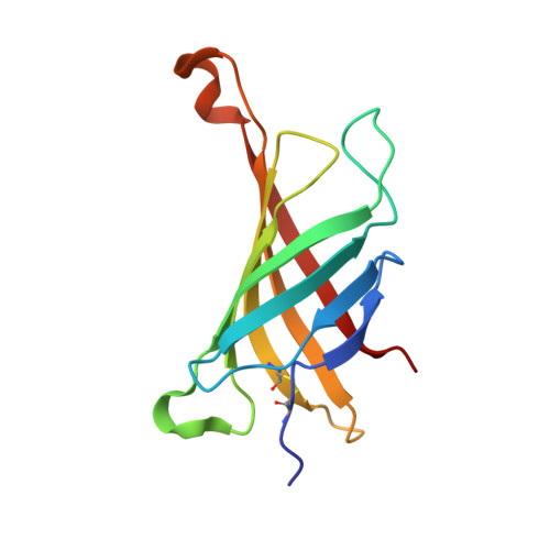
The avidin-theophylline complex: A structural and computational study.

Spinello, A.Lapenta, F.De March, M.

(2023) Proteins 91: 1437-1443

  • DOI: https://doi.org/10.1002/prot.26538
  • Primary Citation of Related Structures:  
    8CK7

  • PubMed Abstract: 

    The interaction between avidin and its counterpart biotin is one of central importance in biology and has been reproposed and studied at length. However, the binding pocket of avidin is prone to promiscuous binding, able to accommodate even non-biotinylated ligands. Comprehending the factors that distinguish the extremely strong interaction with biotin to other ligands is an important step to fully picture the thermodynamics of these low-affinity complexes. Here, we present the complex between chicken white egg avidin and theophylline (TEP), the xanthine derivative used in the therapy of asthma. In the crystal structure, TEP lies in the biotin-binding pocket with the same orientation and planarity of the aromatic ring of 8-oxodeoxyguanosine. Indeed, its affinity for avidin measured by isothermal titration calorimetry is in the same μM range as those obtained for the previously characterized nucleoside derivatives. By the use of molecular dynamic simulations, we have investigated the most important intermolecular interactions occurring in the avidin-TEP binding pocket and compared them with those obtained for the avidin 8-oxodeoxyguanosine and avidin-biotin complexes. These results testify the capability of avidin to complex purely aromatic molecules.


  • Organizational Affiliation

    Department of Biological, Chemical and Pharmaceutical Sciences and Technologies, University of Palermo, Palermo, Italy.


Macromolecules
Find similar proteins by:  (by identity cutoff)  |  3D Structure
Entity ID: 1
MoleculeChains Sequence LengthOrganismDetailsImage
Avidin
A, B
124Gallus gallusMutation(s): 1 
UniProt
Find proteins for P02701 (Gallus gallus)
Explore P02701 
Go to UniProtKB:  P02701
Entity Groups  
Sequence Clusters30% Identity50% Identity70% Identity90% Identity95% Identity100% Identity
UniProt GroupP02701
Sequence Annotations
Expand
  • Reference Sequence
Small Molecules
Ligands 4 Unique
IDChains Name / Formula / InChI Key2D Diagram3D Interactions
NAG
Query on NAG

Download Ideal Coordinates CCD File 
D [auth A],
H [auth B]
2-acetamido-2-deoxy-beta-D-glucopyranose
C8 H15 N O6
OVRNDRQMDRJTHS-FMDGEEDCSA-N
TEP (Subject of Investigation/LOI)
Query on TEP

Download Ideal Coordinates CCD File 
E [auth A],
I [auth B]
THEOPHYLLINE
C7 H8 N4 O2
ZFXYFBGIUFBOJW-UHFFFAOYSA-N
SO4
Query on SO4

Download Ideal Coordinates CCD File 
F [auth A],
G [auth A]
SULFATE ION
O4 S
QAOWNCQODCNURD-UHFFFAOYSA-L
GOL
Query on GOL

Download Ideal Coordinates CCD File 
C [auth A]GLYCEROL
C3 H8 O3
PEDCQBHIVMGVHV-UHFFFAOYSA-N
Experimental Data & Validation

Experimental Data

  • Method: X-RAY DIFFRACTION
  • Resolution: 2.20 Å
  • R-Value Free: 0.233 
  • R-Value Work: 0.184 
  • R-Value Observed: 0.186 
  • Space Group: P 64
Unit Cell:
Length ( Å )Angle ( ˚ )
a = 108.91α = 90
b = 108.91β = 90
c = 43.35γ = 120
Software Package:
Software NamePurpose
REFMACrefinement
iMOSFLMdata reduction
Aimlessdata scaling
MOLREPphasing

Structure Validation

View Full Validation Report


Ligand Structure Quality Assessment 


Entry History & Funding Information

Deposition Data

  • Released Date: 2023-12-27 
  • Deposition Author(s): De March, M.

Funding OrganizationLocationGrant Number
Not funded--

Revision History  (Full details and data files)

  • Version 1.0: 2023-12-27
    Type: Initial release