A Dynamic Water Channel Affects O 2 Stability in [FeFe]-Hydrogenases.
Brocks, C., Das, C.K., Duan, J., Yadav, S., Apfel, U.P., Ghosh, S., Hofmann, E., Winkler, M., Engelbrecht, V., Schafer, L.V., Happe, T.(2024) ChemSusChem 17: e202301365-e202301365
- PubMed: 37830175 Search on PubMed
- DOI: https://doi.org/10.1002/cssc.202301365
- Primary Citation Related Structures:
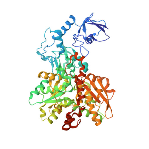
7QHF, 8CJY - PubMed Abstract:
[FeFe]-hydrogenases are capable of reducing protons at a high rate. However, molecular oxygen (O 2 ) induces the degradation of their catalytic cofactor, the H-cluster, which consists of a cubane [4Fe4S] subcluster (4Fe H ) and a unique diiron moiety (2Fe H ). Previous attempts to prevent O 2 -induced damage have focused on enhancing the protein's sieving effect for O 2 by blocking the hydrophobic gas channels that connect the protein surface and the 2Fe H . In this study, we aimed to block an O 2 diffusion pathway and shield 4Fe H instead. Molecular dynamics (MD) simulations identified a novel water channel (W H ) surrounding the H-cluster. As this hydrophilic path may be accessible for O 2 molecules we applied site-directed mutagenesis targeting amino acids along W H in proximity to 4Fe H to block O 2 diffusion. Protein film electrochemistry experiments demonstrate increased O 2 stabilities for variants G302S and S357T, and MD simulations based on high-resolution crystal structures confirmed an enhanced local sieving effect for O 2 in the environment of the 4Fe H in both cases. The results strongly suggest that, in wild type proteins, O 2 diffuses from the 4Fe H to the 2Fe H . These results reveal new strategies for improving the O 2 stability of [FeFe]-hydrogenases by focusing on the O 2 diffusion network near the active site.
- Faculty of Biology and Biotechnology, Photobiotechnology, Ruhr University Bochum, Universitätsstrasse 150, 44801, Bochum, Germany.
Organizational Affiliation: