


Crystal structure of S-adenosyl-L-homocysteine hydrolase from P. aeruginosa in complex with fragment F2X-Entry G03
Malecki, P.H., Gawel, M., Stepniewska, M., Brzezinski, K.To be published.
Experimental Data Snapshot
Starting Model: experimental
View more details
Entity ID: 1 | |||||
|---|---|---|---|---|---|
| Molecule | Chains | Sequence Length | Organism | Details | Image |
| Adenosylhomocysteinase | 472 | Pseudomonas aeruginosa PAO1 | Mutation(s): 0 Gene Names: ahcY, sahH, PA0432 EC: 3.3.1.1 (PDB Primary Data), 3.13.2.1 (UniProt) | 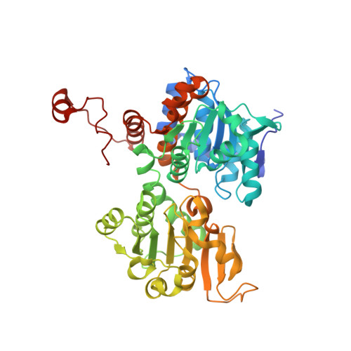 | |
UniProt | |||||
Entity Groups | |||||
| Sequence Clusters | 30% Identity50% Identity70% Identity90% Identity95% Identity100% Identity | ||||
| UniProt Group | Q9I685 | ||||
Sequence AnnotationsExpand | |||||
Reference Sequence | |||||
| Ligands 6 Unique | |||||
|---|---|---|---|---|---|
| ID | Chains | Name / Formula / InChI Key | 2D Diagram | 3D Interactions | |
| NAD Download:Ideal Coordinates CCD File | BA [auth D], E [auth A], M [auth B], T [auth C] | NICOTINAMIDE-ADENINE-DINUCLEOTIDE C21 H27 N7 O14 P2 BAWFJGJZGIEFAR-NNYOXOHSSA-N |  | ||
| RD4 (Subject of Investigation/LOI) Download:Ideal Coordinates CCD File | G [auth A], L [auth B], S [auth C] | 3-ethoxybenzene-1-carboximidamide C9 H12 N2 O OZPMVTBYBADRPR-UHFFFAOYSA-N |  | ||
| ADE Download:Ideal Coordinates CCD File | CA [auth D], F [auth A], N [auth B], U [auth C] | ADENINE C5 H5 N5 GFFGJBXGBJISGV-UHFFFAOYSA-N |  | ||
| PO4 Download:Ideal Coordinates CCD File | AA [auth C] EA [auth D] FA [auth D] I [auth A] J [auth A] | PHOSPHATE ION O4 P NBIIXXVUZAFLBC-UHFFFAOYSA-K |  | ||
| DMS Download:Ideal Coordinates CCD File | V [auth C] | DIMETHYL SULFOXIDE C2 H6 O S IAZDPXIOMUYVGZ-UHFFFAOYSA-N |  | ||
| K Download:Ideal Coordinates CCD File | DA [auth D], H [auth A], O [auth B], W [auth C] | POTASSIUM ION K NPYPAHLBTDXSSS-UHFFFAOYSA-N |  | ||
| Length ( Å ) | Angle ( ˚ ) |
|---|---|
| a = 91.208 | α = 90 |
| b = 101.49 | β = 114.42 |
| c = 110.704 | γ = 90 |
| Software Name | Purpose |
|---|---|
| PHENIX | refinement |
| XDS | data scaling |
| XDS | data reduction |
| REFMAC | phasing |
| Funding Organization | Location | Grant Number |
|---|---|---|
| Polish National Science Centre | Poland | SONATA BIS 2018/30/E/NZ1/00729 |


