Biochemical and structural characterisation of a family GH5 cellulase from endosymbiont of shipworm P. megotara.
Junghare, M., Manavalan, T., Fredriksen, L., Leiros, I., Altermark, B., Eijsink, V.G.H., Vaaje-Kolstad, G.(2023) Biotechnol Biofuels Bioprod 16: 61-61
- PubMed: 37016457 Search on PubMedSearch on PubMed Central
- DOI: https://doi.org/10.1186/s13068-023-02307-1
- Primary Citation Related Structures:
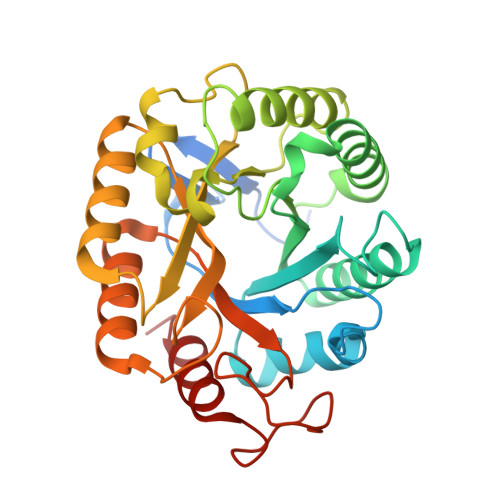
8C10 - PubMed Abstract:
Cellulases play a key role in the enzymatic conversion of plant cell-wall polysaccharides into simple and economically relevant sugars. Thus, the discovery of novel cellulases from exotic biological niches is of great interest as they may present properties that are valuable in the biorefining of lignocellulosic biomass. We have characterized a glycoside hydrolase 5 (GH5) domain of a bi-catalytic GH5-GH6 multi-domain enzyme from the unusual gill endosymbiont Teredinibacter waterburyi of the wood-digesting shipworm Psiloteredo megotara. The catalytic GH5 domain, was cloned and recombinantly produced with or without a C-terminal family 10 carbohydrate-binding module (CBM). Both variants showed hydrolytic endo-activity on soluble substrates such as β-glucan, carboxymethylcellulose and konjac glucomannan, respectively. However, low activity was observed towards the crystalline form of cellulose. Interestingly, when co-incubated with a cellulose-active LPMO, a clear synergy was observed that boosted the overall hydrolysis of crystalline cellulose. The crystal structure of the GH5 catalytic domain was solved to 1.0 Å resolution and revealed a substrate binding cleft extension containing a putative + 3 subsite, which is uncommon in this enzyme family. The enzyme was active in a wide range of pH, temperatures and showed high tolerance for NaCl. This study provides significant knowledge in the discovery of new enzymes from shipworm gill endosymbionts and sheds new light on biochemical and structural characterization of cellulolytic cellulase. Study demonstrated a boost in the hydrolytic activity of cellulase on crystalline cellulose when co-incubated with cellulose-active LPMO. These findings will be relevant for the development of future enzyme cocktails that may be useful for the biotechnological conversion of lignocellulose.
- Department of Chemistry, Biotechnology and Food Science, Norwegian University of Life Sciences, 1432, Ås, Norway. madan.junghare@nmbu.no.
Organizational Affiliation: