Molecular basis of JAK2 activation in erythropoietin receptor and pathogenic JAK2 signaling.
Abraham, B.G., Haikarainen, T., Vuorio, J., Girych, M., Virtanen, A.T., Kurttila, A., Karathanasis, C., Heilemann, M., Sharma, V., Vattulainen, I., Silvennoinen, O.(2024) Sci Adv 10: eadl2097-eadl2097
- PubMed: 38457493 Search on PubMedSearch on PubMed Central
- DOI: https://doi.org/10.1126/sciadv.adl2097
- Primary Citation Related Structures:
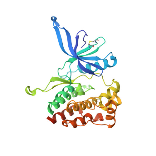
8C08, 8C09, 8C0A - PubMed Abstract:
Janus kinase 2 (JAK2) mediates type I/II cytokine receptor signaling, but JAK2 is also activated by somatic mutations that cause hematological malignancies by mechanisms that are still incompletely understood. Quantitative superresolution microscopy (qSMLM) showed that erythropoietin receptor (EpoR) exists as monomers and dimerizes upon Epo stimulation or through the predominant JAK2 pseudokinase domain mutations (V617F, K539L, and R683S). Crystallographic analysis complemented by kinase activity analysis and atomic-level simulations revealed distinct pseudokinase dimer interfaces and activation mechanisms for the mutants: JAK V617F activity is driven by dimerization, K539L involves both increased receptor dimerization and kinase activity, and R683S prevents autoinhibition and increases catalytic activity and drives JAK2 equilibrium toward activation state through a wild-type dimer interface. Artificial intelligence-guided modeling and simulations revealed that the pseudokinase mutations cause differences in the pathogenic full-length JAK2 dimers, particularly in the FERM-SH2 domains. A detailed molecular understanding of mutation-driven JAK2 hyperactivation may enable novel therapeutic approaches to selectively target pathogenic JAK2 signaling.
- Faculty of Medicine and Health Technology, Tampere University, Tampere, Finland.
Organizational Affiliation: