8BW0 | pdb_00008bw0

Structure of CEACAM5 A3-B3 domain in Complex with Tusamitamab Fab


Experimental Data Snapshot

  • Method: ELECTRON MICROSCOPY
  • Resolution: 3.11 Å
  • Aggregation State: PARTICLE 
  • Reconstruction Method: SINGLE PARTICLE 

wwPDB Validation   3D Report Full Report


This is version 1.2 of the entry. See complete history


Literature

Structural insights into epitope-paratope interactions of a monoclonal antibody targeting CEACAM5-expressing tumors.

Kumar, A.Duffieux, F.Gagnaire, M.Rapisarda, C.Bertrand, T.Rak, A.

(2024) Nat Commun 15: 9377-9377

  • DOI: https://doi.org/10.1038/s41467-024-53746-9
  • Primary Citation of Related Structures:  
    8BW0

  • PubMed Abstract: 

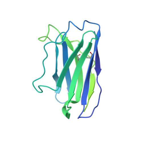
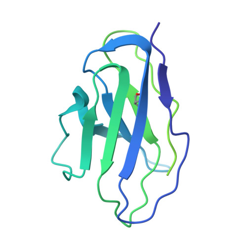
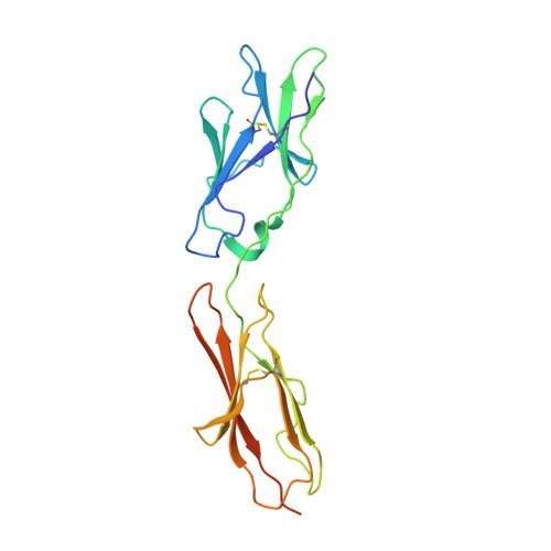
    Carcinoembryonic antigen-related cell adhesion molecules (CEACAMs) are overexpressed in some tumor types. The antibody-drug conjugate tusamitamab ravtansine specifically recognizes the A3-B3 domains of human CEACAM5 (hCEACAM5). To understand this specificity, here we map the epitope-paratope interface between the A3-B3 domains of hCEACAM5 (hCEACAM5 A3-B3 ) and the antigen-binding fragment of tusamitamab (tusa Fab). We use hydrogen/deuterium exchange mass spectrometry to identify the tusa Fab paratope, which involves heavy chain (HC) residues 101-109 and light chain residues 48-54 and 88-104. Using surface plasmon resonance, we demonstrate that alanine variants of HC residues 96-108 abolish binding to hCEACAM5, suggesting that these residues are critical for tusa-Fab-antigen complex formation. The cryogenic electron microscopy structure of the hCEACAM5 A3-B3 - tusa Fab complex (3.11 Å overall resolution) reveals a discontinuous epitope involving residues in the A3-B3 domains and an N-linked mannose at residue Asn612. Conformational constraints on the epitope-paratope interface enable tusamitamab to target hCEACAM5 A3-B3 and distinguish CEACAM5 from other CEACAMs.


  • Organizational Affiliation
    • Integrated Drug Discovery, Sanofi R&D, Paris, France.

Macromolecules
Find similar proteins by:  (by identity cutoff)  |  3D Structure
Entity ID: 1
MoleculeChains Sequence LengthOrganismDetailsImage
Tusamitamab Fab heavy ChainA [auth H]234Mus sp.Mutation(s): 0 
Entity Groups  
Sequence Clusters30% Identity50% Identity70% Identity90% Identity95% Identity100% Identity
Sequence Annotations
Expand
  • Reference Sequence
Find similar proteins by:  (by identity cutoff)  |  3D Structure
Entity ID: 2
MoleculeChains Sequence LengthOrganismDetailsImage
Tusamitamab Light ChainB [auth L]214Mus sp.Mutation(s): 0 
Entity Groups  
Sequence Clusters30% Identity50% Identity70% Identity90% Identity95% Identity100% Identity
Sequence Annotations
Expand
  • Reference Sequence
Find similar proteins by:  (by identity cutoff)  |  3D Structure
Entity ID: 3
MoleculeChains Sequence LengthOrganismDetailsImage
Carcinoembryonic antigen-related cell adhesion molecule 5193Homo sapiensMutation(s): 0 
Gene Names: CEACAM5CEA
UniProt & NIH Common Fund Data Resources
Find proteins for P06731 (Homo sapiens)
Explore P06731 
Go to UniProtKB:  P06731
PHAROS:  P06731
GTEx:  ENSG00000105388 
Entity Groups  
Sequence Clusters30% Identity50% Identity70% Identity90% Identity95% Identity100% Identity
UniProt GroupP06731
Glycosylation
Glycosylation Sites: 6Go to GlyGen: P06731-1
Sequence Annotations
Expand
  • Reference Sequence
Oligosaccharides

Help

Entity ID: 4
MoleculeChains Length2D Diagram Glycosylation3D Interactions
2-acetamido-2-deoxy-beta-D-glucopyranose-(1-4)-2-acetamido-2-deoxy-beta-D-glucopyranoseD [auth A],
F [auth D]
2N-Glycosylation
Glycosylation Resources
GlyTouCan:  G42666HT
GlyCosmos:  G42666HT
GlyGen:  G42666HT
Entity ID: 5
MoleculeChains Length2D Diagram Glycosylation3D Interactions
alpha-D-mannopyranose-(1-3)-[alpha-D-mannopyranose-(1-6)]beta-D-mannopyranose-(1-4)-2-acetamido-2-deoxy-beta-D-glucopyranose-(1-4)-[alpha-L-fucopyranose-(1-6)]2-acetamido-2-deoxy-beta-D-glucopyranoseE [auth B]6N-Glycosylation
Glycosylation Resources
GlyTouCan:  G82348BZ
GlyCosmos:  G82348BZ
GlyGen:  G82348BZ
Experimental Data & Validation

Experimental Data

  • Method: ELECTRON MICROSCOPY
  • Resolution: 3.11 Å
  • Aggregation State: PARTICLE 
  • Reconstruction Method: SINGLE PARTICLE 
EM Software:
TaskSoftware PackageVersion
RECONSTRUCTIONcryoSPARC3.2
MODEL REFINEMENTPHENIX1.19.2

Structure Validation

View Full Validation Report


Entry History & Funding Information

Deposition Data


Funding OrganizationLocationGrant Number
Other privateFrance--

Revision History  (Full details and data files)

  • Version 1.0: 2024-01-24
    Type: Initial release
  • Version 1.1: 2024-10-16
    Changes: Data collection, Structure summary
  • Version 1.2: 2024-11-13
    Changes: Data collection, Database references