Structural and Functional Characterization of the Ureidoacrylate Amidohydrolase RutB from Escherichia coli .
Busch, M.R., Rajendran, C., Sterner, R.(2023) Biochemistry 62: 863-872
- PubMed: 36599150 Search on PubMed
- DOI: https://doi.org/10.1021/acs.biochem.2c00640
- Primary Citation Related Structures:
8BKD, 8BLL, 8BLM, 8BLN, 8BYW - PubMed Abstract:
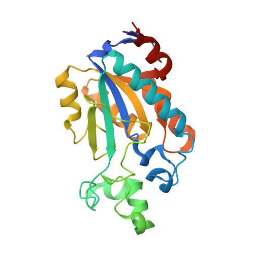
We present a detailed structure-function analysis of the ureidoacrylate amidohydrolase RutB from Eschericha coli , which is an essential enzyme of the Rut pathway for pyrimidine utilization. Crystals of selenomethionine-labeled RutB were produced, which allowed us to determine the first structure of the enzyme at a resolution of 1.9 Å and to identify it as a new member of the isochorismatase-like hydrolase family. RutB was co-crystallized with the substrate analogue ureidopropionate, revealing the mode of substrate binding. Mutation of residues constituting the catalytic triad (D24A, D24N, K133A, C166A, C166S, C166T, C166Y) resulted in complete inactivation of RutB, whereas mutation of other residues close to the active site (Y29F, Y35F, N72A, W74A, W74F, E80A, E80D, S92A, S92T, S92Y, Q105A, Y136A, Y136F) leads to distinct changes of the turnover number ( k cat ) and/or the Michaelis constant ( K M ). The results of our structural and mutational studies allowed us to assign specific functions to individual residues and to formulate a plausible reaction mechanism for RutB.
- Institute of Biophysics and Physical Biochemistry, Regensburg Center for Biochemistry, University of Regensburg, D-93040 Regensburg, Germany.
Organizational Affiliation: