DNA-origami-directed virus capsid polymorphism.
Seitz, I., Saarinen, S., Kumpula, E.P., McNeale, D., Anaya-Plaza, E., Lampinen, V., Hytonen, V.P., Sainsbury, F., Cornelissen, J.J.L.M., Linko, V., Huiskonen, J.T., Kostiainen, M.A.(2023) Nat Nanotechnol 18: 1205-1212
- PubMed: 37460794 Search on PubMedSearch on PubMed Central
- DOI: https://doi.org/10.1038/s41565-023-01443-x
- Primary Citation Related Structures:
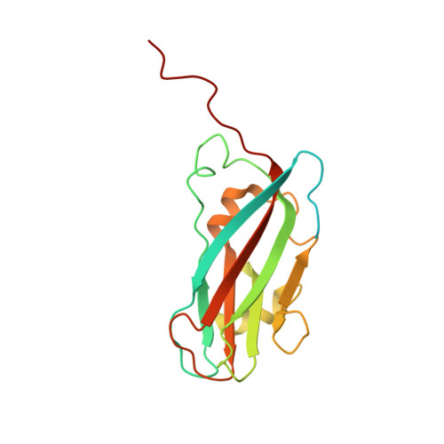
8BI4 - PubMed Abstract:
Viral capsids can adopt various geometries, most iconically characterized by icosahedral or helical symmetries. Importantly, precise control over the size and shape of virus capsids would have advantages in the development of new vaccines and delivery systems. However, current tools to direct the assembly process in a programmable manner are exceedingly elusive. Here we introduce a modular approach by demonstrating DNA-origami-directed polymorphism of single-protein subunit capsids. We achieve control over the capsid shape, size and topology by employing user-defined DNA origami nanostructures as binding and assembly platforms, which are efficiently encapsulated within the capsid. Furthermore, the obtained viral capsid coatings can shield the encapsulated DNA origami from degradation. Our approach is, moreover, not limited to a single type of capsomers and can also be applied to RNA-DNA origami structures to pave way for next-generation cargo protection and targeting strategies.
- Department of Bioproducts and Biosystems, Aalto University, Aalto, Finland.
Organizational Affiliation: