Diffraction anisotropy and paired refinement: crystal structure of H33, a protein binder to interleukin 10.
Kolenko, P., Mikulecky, P., Pham, P.N., Maly, M., Schneider, B.(2023) J Appl Crystallogr 56: 1261-1266
- PubMed: 37555209 Search on PubMedSearch on PubMed Central
- DOI: https://doi.org/10.1107/S160057672300479X
- Primary Citation Related Structures:
8BDU - PubMed Abstract:
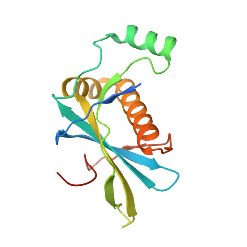
Binder H33 is a small protein binder engineered by ribosome display to bind human interleukin 10. Crystals of binder H33 display severe diffraction anisotropy. A set of data files with correction for diffraction anisotropy based on different local signal-to-noise ratios was prepared. Paired refinement was used to find the optimal anisotropic high-resolution diffraction limit of the data: 3.13-2.47 Å. The structure of binder H33 belongs to the 2% of crystal structures with the highest solvent content in the Protein Data Bank.
- Czech Technical University in Prague, Brehova 7, Prague 115 19, Czech Republic.
Organizational Affiliation: