8BD4

TniQ-capped Tns-ATP-dsDNA complex


Experimental Data Snapshot

  • Method: ELECTRON MICROSCOPY
  • Resolution: 3.44 Å
  • Aggregation State: PARTICLE 
  • Reconstruction Method: SINGLE PARTICLE 

wwPDB Validation   3D Report Full Report


This is version 1.1 of the entry. See complete history


Literature

Structural basis for the assembly of the type V CRISPR-associated transposon complex.

Schmitz, M.Querques, I.Oberli, S.Chanez, C.Jinek, M.

(2022) Cell 185: 4999

  • DOI: https://doi.org/10.1016/j.cell.2022.11.009
  • Primary Citation of Related Structures:  
    8BD4, 8BD5, 8BD6

  • PubMed Abstract: 

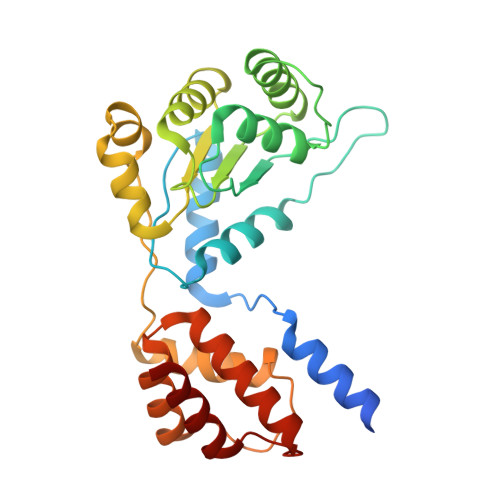
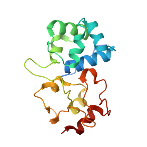
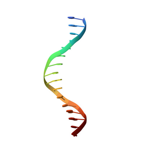
    CRISPR-Cas systems have been co-opted by Tn7-like transposable elements to direct RNA-guided transposition. Type V-K CRISPR-associated transposons rely on the concerted activities of the pseudonuclease Cas12k, the AAA+ ATPase TnsC, the Zn-finger protein TniQ, and the transposase TnsB. Here we present a cryo-electron microscopic structure of a target DNA-bound Cas12k-transposon recruitment complex comprised of RNA-guided Cas12k, TniQ, a polymeric TnsC filament and, unexpectedly, the ribosomal protein S15. Complex assembly, mediated by a network of interactions involving the guide RNA, TniQ, and S15, results in R-loop completion. TniQ contacts two TnsC protomers at the Cas12k-proximal filament end, likely nucleating its polymerization. Transposition activity assays corroborate our structural findings, implying that S15 is a bona fide component of the type V crRNA-guided transposon machinery. Altogether, our work uncovers key mechanistic aspects underpinning RNA-mediated assembly of CRISPR-associated transposons to guide their development as programmable tools for site-specific insertion of large DNA payloads.


  • Organizational Affiliation

    Department of Biochemistry, University of Zurich, Zurich 8057, Switzerland.


Macromolecules

Find similar proteins by:  (by identity cutoff)  |  3D Structure
Entity ID: 1
MoleculeChains Sequence LengthOrganismDetailsImage
TnsC
A, B, C, D, E
A, B, C, D, E, F, G
276Scytonema hofmanniiMutation(s): 0 
UniProt
Find proteins for A0A8J0PCL3 (Scytonema hofmannii)
Explore A0A8J0PCL3 
Go to UniProtKB:  A0A8J0PCL3
Entity Groups  
Sequence Clusters30% Identity50% Identity70% Identity90% Identity95% Identity100% Identity
UniProt GroupA0A8J0PCL3
Sequence Annotations
Expand
  • Reference Sequence
Find similar proteins by:  (by identity cutoff)  |  3D Structure
Entity ID: 2
MoleculeChains Sequence LengthOrganismDetailsImage
TniQ (Homology model)H [auth R],
I [auth S],
J [auth T]
167Scytonema hofmanniiMutation(s): 0 
UniProt
Find proteins for A0A8J0PCL5 (Scytonema hofmannii)
Explore A0A8J0PCL5 
Go to UniProtKB:  A0A8J0PCL5
Entity Groups  
Sequence Clusters30% Identity50% Identity70% Identity90% Identity95% Identity100% Identity
UniProt GroupA0A8J0PCL5
Sequence Annotations
Expand
  • Reference Sequence

Find similar nucleic acids by:  Sequence   |   3D Structure  

Entity ID: 3
MoleculeChains LengthOrganismImage
DNA (5'-D(P*GP*AP*TP*CP*GP*AP*TP*CP*GP*AP*TP*CP*GP*AP*TP*C)-3')K [auth U],
L [auth V]
16synthetic construct
Sequence Annotations
Expand
  • Reference Sequence
Small Molecules
Ligands 3 Unique
IDChains Name / Formula / InChI Key2D Diagram3D Interactions
ATP (Subject of Investigation/LOI)
Query on ATP

Download Ideal Coordinates CCD File 
AA [auth G]
M [auth A]
P [auth B]
R [auth C]
T [auth D]
AA [auth G],
M [auth A],
P [auth B],
R [auth C],
T [auth D],
V [auth E],
Y [auth F]
ADENOSINE-5'-TRIPHOSPHATE
C10 H16 N5 O13 P3
ZKHQWZAMYRWXGA-KQYNXXCUSA-N
ZN (Subject of Investigation/LOI)
Query on ZN

Download Ideal Coordinates CCD File 
CA [auth R]
DA [auth S]
EA [auth T]
FA [auth T]
O [auth A]
CA [auth R],
DA [auth S],
EA [auth T],
FA [auth T],
O [auth A],
X [auth E]
ZINC ION
Zn
PTFCDOFLOPIGGS-UHFFFAOYSA-N
MG (Subject of Investigation/LOI)
Query on MG

Download Ideal Coordinates CCD File 
BA [auth G]
N [auth A]
Q [auth B]
S [auth C]
U [auth D]
BA [auth G],
N [auth A],
Q [auth B],
S [auth C],
U [auth D],
W [auth E],
Z [auth F]
MAGNESIUM ION
Mg
JLVVSXFLKOJNIY-UHFFFAOYSA-N
Experimental Data & Validation

Experimental Data

  • Method: ELECTRON MICROSCOPY
  • Resolution: 3.44 Å
  • Aggregation State: PARTICLE 
  • Reconstruction Method: SINGLE PARTICLE 

Structure Validation

View Full Validation Report


Entry History & Funding Information

Deposition Data


Funding OrganizationLocationGrant Number
European Research Council (ERC)European Union820152
Swiss National Science FoundationSwitzerland31003A_182567
European Molecular Biology Organization (EMBO)European UnionALTF 296-2020
Howard Hughes Medical Institute (HHMI)United States55008735

Revision History  (Full details and data files)

  • Version 1.0: 2022-12-28
    Type: Initial release
  • Version 1.1: 2023-01-04
    Changes: Database references