8BCA

Human Brr2 Helicase Region in complex with C-tail deleted Jab1 and compound 26


Experimental Data Snapshot

  • Method: X-RAY DIFFRACTION
  • Resolution: 2.80 Å
  • R-Value Free: 0.258 
  • R-Value Work: 0.199 
  • R-Value Observed: 0.201 

wwPDB Validation   3D Report Full Report


Ligand Structure Quality Assessment 


This is version 1.2 of the entry. See complete history


Literature

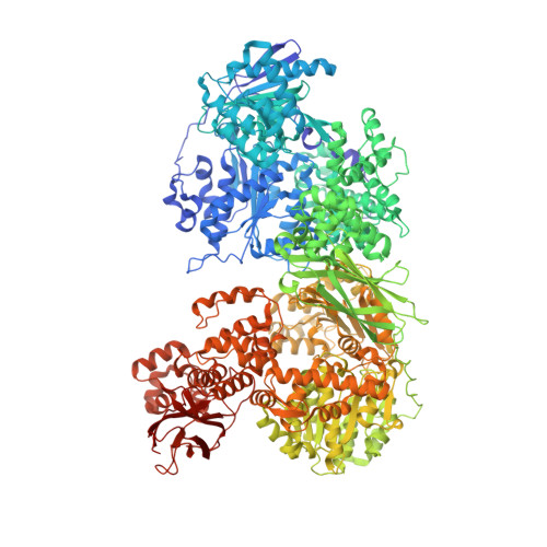
Conformation-dependent ligand hot spots in the spliceosomal RNA helicase BRR2.

Vester, K.Metz, A.Huber, S.Loll, B.Wahl, M.C.

(2023) Acta Crystallogr D Struct Biol 79: 304-317

  • DOI: https://doi.org/10.1107/S2059798323001778
  • Primary Citation of Related Structures:  
    8BC8, 8BC9, 8BCA, 8BCB, 8BCC, 8BCD, 8BCE, 8BCF, 8BCG, 8BCH

  • PubMed Abstract: 

    The conversion of hits to leads in drug discovery involves the elaboration of chemical core structures to increase their potency. In fragment-based drug discovery, low-molecular-weight compounds are tested for protein binding and are subsequently modified, with the tacit assumption that the binding mode of the original hit will be conserved among the derivatives. However, deviations from binding mode conservation are rather frequently observed, but potential causes of these alterations remain incompletely understood. Here, two crystal forms of the spliceosomal RNA helicase BRR2 were employed as a test case to explore the consequences of conformational changes in the target protein on the binding behaviour of fragment derivatives. The initial fragment, sulfaguanidine, bound at the interface between the two helicase cassettes of BRR2 in one crystal form. Second-generation compounds devised by structure-guided docking were probed for their binding to BRR2 in a second crystal form, in which the original fragment-binding site was altered due to a conformational change. While some of the second-generation compounds retained binding to parts of the original site, others changed to different binding pockets of the protein. A structural bioinformatics analysis revealed that the fragment-binding sites correspond to predicted binding hot spots, which strongly depend on the protein conformation. This case study offers an example of extensive binding-mode changes during hit derivatization, which are likely to occur as a consequence of multiple binding hot spots, some of which are sensitive to the flexibility of the protein.


  • Organizational Affiliation

    Laboratory of Structural Biochemistry, Institute of Chemistry and Biochemistry, Freie Universität Berlin, Takustrasse 6, 14195 Berlin, Germany.


Macromolecules
Find similar proteins by:  (by identity cutoff)  |  3D Structure
Entity ID: 1
MoleculeChains Sequence LengthOrganismDetailsImage
U5 small nuclear ribonucleoprotein 200 kDa helicaseA [auth B]1,747Homo sapiensMutation(s): 0 
Gene Names: SNRNP200ASCC3L1HELIC2KIAA0788
EC: 3.6.4.13
UniProt & NIH Common Fund Data Resources
Find proteins for O75643 (Homo sapiens)
Explore O75643 
Go to UniProtKB:  O75643
PHAROS:  O75643
GTEx:  ENSG00000144028 
Entity Groups  
Sequence Clusters30% Identity50% Identity70% Identity90% Identity95% Identity100% Identity
UniProt GroupO75643
Sequence Annotations
Expand
  • Reference Sequence
Find similar proteins by:  (by identity cutoff)  |  3D Structure
Entity ID: 2
MoleculeChains Sequence LengthOrganismDetailsImage
Pre-mRNA-processing-splicing factor 8B [auth J]263Homo sapiensMutation(s): 0 
Gene Names: PRPF8PRPC8
UniProt & NIH Common Fund Data Resources
Find proteins for Q6P2Q9 (Homo sapiens)
Explore Q6P2Q9 
Go to UniProtKB:  Q6P2Q9
PHAROS:  Q6P2Q9
GTEx:  ENSG00000174231 
Entity Groups  
Sequence Clusters30% Identity50% Identity70% Identity90% Identity95% Identity100% Identity
UniProt GroupQ6P2Q9
Sequence Annotations
Expand
  • Reference Sequence
Small Molecules
Ligands 3 Unique
IDChains Name / Formula / InChI Key2D Diagram3D Interactions
Q96 (Subject of Investigation/LOI)
Query on Q96

Download Ideal Coordinates CCD File 
C [auth B],
D [auth B]
3-azanyl-~{N}-methyl-4-(methylamino)benzenesulfonamide
C8 H13 N3 O2 S
BSCVJYVFYUNMQC-UHFFFAOYSA-N
GOL
Query on GOL

Download Ideal Coordinates CCD File 
F [auth B]GLYCEROL
C3 H8 O3
PEDCQBHIVMGVHV-UHFFFAOYSA-N
EDO
Query on EDO

Download Ideal Coordinates CCD File 
E [auth B]
G [auth B]
H [auth B]
I [auth B]
J [auth B]
E [auth B],
G [auth B],
H [auth B],
I [auth B],
J [auth B],
K [auth B],
L [auth B],
M [auth B],
N [auth B]
1,2-ETHANEDIOL
C2 H6 O2
LYCAIKOWRPUZTN-UHFFFAOYSA-N
Experimental Data & Validation

Experimental Data

  • Method: X-RAY DIFFRACTION
  • Resolution: 2.80 Å
  • R-Value Free: 0.258 
  • R-Value Work: 0.199 
  • R-Value Observed: 0.201 
  • Space Group: P 21 21 21
Unit Cell:
Length ( Å )Angle ( ˚ )
a = 100.409α = 90
b = 119.362β = 90
c = 187.765γ = 90
Software Package:
Software NamePurpose
PHENIXrefinement
XDSdata reduction
XSCALEdata scaling
PHASERphasing

Structure Validation

View Full Validation Report


Ligand Structure Quality Assessment 


Entry History & Funding Information

Deposition Data


Funding OrganizationLocationGrant Number
German Research Foundation (DFG)Germany278001972

Revision History  (Full details and data files)

  • Version 1.0: 2023-03-08
    Type: Initial release
  • Version 1.1: 2023-04-19
    Changes: Database references
  • Version 1.2: 2024-02-07
    Changes: Data collection, Refinement description