Evaluation of Auranofin Loading within Ferritin Nanocages.
Lucignano, R., Pratesi, A., Imbimbo, P., Monti, D.M., Picone, D., Messori, L., Ferraro, G., Merlino, A.(2022) Int J Mol Sci 23
- PubMed: 36430642 Search on PubMedSearch on PubMed Central
- DOI: https://doi.org/10.3390/ijms232214162
- Primary Citation Related Structures:
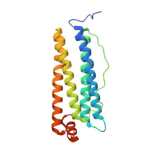
8B7L, 8B7O - PubMed Abstract:
Auranofin (AF), a gold(I) compound that is currently used for the treatment of rheumatoid arthritis and is in clinical trials for its promising anticancer activity, was encapsulated within the human H-chain and the horse spleen ferritin nanocages using the alkaline disassembly/reassembly protocol. The aim of the work was to highlight possible differences in their drug loading capacity and efficacy. The drug-loaded ferritins were characterized via UV-vis absorption spectroscopy and inductively coupled plasma-atomic emission spectroscopy to assess AF encapsulation and to define the exact amount of gold atoms trapped in the Ft cavity. The crystal structures allowed us to define the nature of AF interaction with both ferritins and to identify the gold binding sites. Moreover, the biological characterization let us to obtain preliminary information on the cytotoxic effect of AF when bound to the human H-chain.
- Department of Chemical Sciences, University of Naples Federico II, Complesso Universitario di Monte Sant'Angelo, Via Cinthia 21, 80126 Naples, Italy.
Organizational Affiliation: