8B77

The crystal structure of N828V variant of DNA Pol Epsilon containing dATP in the polymerase active site


Experimental Data Snapshot

  • Method: X-RAY DIFFRACTION
  • Resolution: 2.70 Å
  • R-Value Free: 0.273 
  • R-Value Work: 0.236 
  • R-Value Observed: 0.238 

wwPDB Validation   3D Report Full Report


Ligand Structure Quality Assessment 


This is version 1.1 of the entry. See complete history


Literature

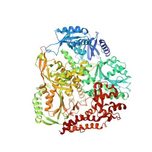
A sensor complements the steric gate when DNA polymerase epsilon discriminates ribonucleotides.

Parkash, V.Kulkarni, Y.Bylund, G.O.Osterman, P.Kamerlin, S.C.L.Johansson, E.

(2023) Nucleic Acids Res 

  • DOI: https://doi.org/10.1093/nar/gkad817
  • Primary Citation of Related Structures:  
    8B67, 8B6K, 8B76, 8B77, 8B79, 8B7E

  • PubMed Abstract: 

    The cellular imbalance between high concentrations of ribonucleotides (NTPs) and low concentrations of deoxyribonucleotides (dNTPs), is challenging for DNA polymerases when building DNA from dNTPs. It is currently believed that DNA polymerases discriminate against NTPs through a steric gate model involving a clash between a tyrosine and the 2'-hydroxyl of the ribonucleotide in the polymerase active site in B-family DNA polymerases. With the help of crystal structures of a B-family polymerase with a UTP or CTP in the active site, molecular dynamics simulations, biochemical assays and yeast genetics, we have identified a mechanism by which the finger domain of the polymerase sense NTPs in the polymerase active site. In contrast to the previously proposed polar filter, our experiments suggest that the amino acid residue in the finger domain senses ribonucleotides by steric hindrance. Furthermore, our results demonstrate that the steric gate in the palm domain and the sensor in the finger domain are both important when discriminating NTPs. Structural comparisons reveal that the sensor residue is conserved among B-family polymerases and we hypothesize that a sensor in the finger domain should be considered in all types of DNA polymerases.


  • Organizational Affiliation

    Department of Medical Biochemistry and Biophysics, Umeå University, Umeå 90187, Sweden.


Macromolecules

Find similar proteins by:  (by identity cutoff)  |  3D Structure
Entity ID: 1
MoleculeChains Sequence LengthOrganismDetailsImage
DNA polymerase epsilon catalytic subunit A1,191Saccharomyces cerevisiaeMutation(s): 3 
Gene Names: POL2DUN2YNL262WN0825
EC: 2.7.7.7 (PDB Primary Data), 3.1.11 (PDB Primary Data)
UniProt
Find proteins for P21951 (Saccharomyces cerevisiae (strain ATCC 204508 / S288c))
Explore P21951 
Go to UniProtKB:  P21951
Entity Groups  
Sequence Clusters30% Identity50% Identity70% Identity90% Identity95% Identity100% Identity
UniProt GroupP21951
Sequence Annotations
Expand
  • Reference Sequence

Find similar nucleic acids by:  Sequence   |   3D Structure  

Entity ID: 2
MoleculeChains LengthOrganismImage
Primer DNA sequenceB [auth P]11synthetic construct
Sequence Annotations
Expand
  • Reference Sequence

Find similar nucleic acids by:  Sequence   |   3D Structure  

Entity ID: 3
MoleculeChains LengthOrganismImage
Template DNA sequenceC [auth T]16synthetic construct
Sequence Annotations
Expand
  • Reference Sequence
Small Molecules
Experimental Data & Validation

Experimental Data

  • Method: X-RAY DIFFRACTION
  • Resolution: 2.70 Å
  • R-Value Free: 0.273 
  • R-Value Work: 0.236 
  • R-Value Observed: 0.238 
  • Space Group: C 1 2 1
Unit Cell:
Length ( Å )Angle ( ˚ )
a = 158.994α = 90
b = 71.03β = 112.99
c = 155.255γ = 90
Software Package:
Software NamePurpose
PHENIXrefinement
PDB_EXTRACTdata extraction
XDSdata reduction
XSCALEdata scaling
PHASERphasing

Structure Validation

View Full Validation Report


Ligand Structure Quality Assessment 


Entry History & Funding Information

Deposition Data


Funding OrganizationLocationGrant Number
CancerfondenSweden--
Swedish Research CouncilSweden--

Revision History  (Full details and data files)

  • Version 1.0: 2023-10-25
    Type: Initial release
  • Version 1.1: 2023-11-01
    Changes: Database references