Exploring the Ligand Binding and Conformational Dynamics of the Substrate-Binding Domain 1 of the ABC Transporter GlnPQ.
Nemchinova, M., Schuurman-Wolters, G.K., Whittaker, J.J., Arkhipova, V., Marrink, S.J., Poolman, B., Guskov, A.(2024) J Phys Chem B 128: 7822-7832
- PubMed: 39090964 Search on PubMedSearch on PubMed Central
- DOI: https://doi.org/10.1021/acs.jpcb.4c02662
- Primary Citation Related Structures:
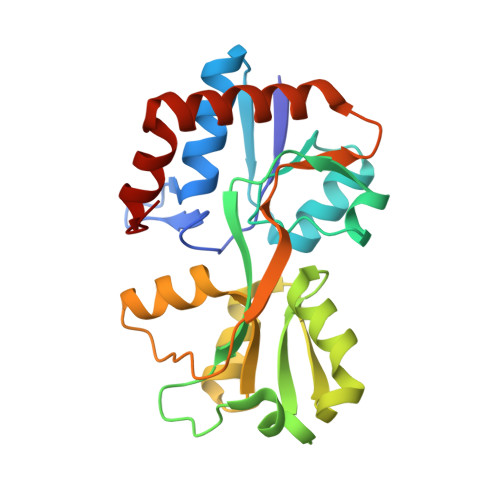
8B5D, 8B5E - PubMed Abstract:
The adenosine triphosphate (ATP)-binding cassette (ABC) importer GlnPQ from Lactococcus lactis has two sequential covalently linked substrate-binding domains (SBDs), which capture the substrates and deliver them to the translocon. The two SBDs differ in their ligand specificities, binding affinities and the distance to the transmembrane domain; interestingly, both SBDs can bind their ligands simultaneously without affecting each other. In this work, we studied the binding of ligands to both SBDs using X-ray crystallography and molecular dynamics simulations. We report three high-resolution structures of SBD1, namely, the wild-type SBD1 with bound asparagine or arginine, and E184D SBD1 with glutamine bound. Molecular dynamics (MD) simulations provide a detailed insight into the dynamics associated with open-closed transitions of the SBDs.
- Groningen Institute for Biomolecular Sciences and Biotechnology, University of Groningen, 9747AG Groningen, The Netherlands.
Organizational Affiliation: