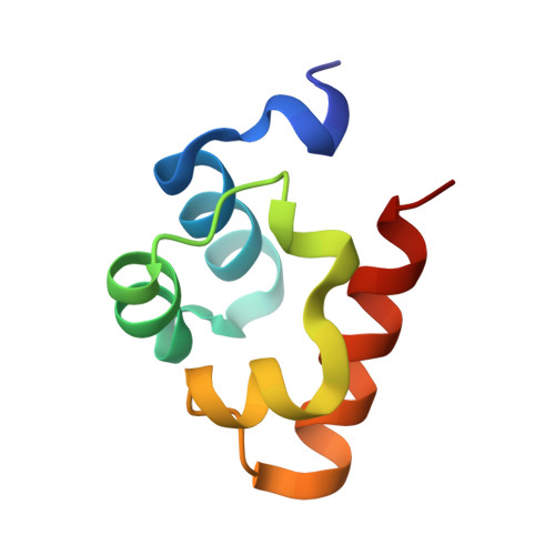
Structural deficits in key domains of Shank2 lead to alterations in postsynaptic nanoclusters and to a neurodevelopmental disorder in humans.
Hassani Nia, F., Woike, D., Bento, I., Niebling, S., Tibbe, D., Schulz, K., Hirnet, D., Skiba, M., Honck, H.H., Veith, K., Gunther, C., Scholz, T., Bierhals, T., Driemeyer, J., Bend, R., Failla, A.V., Lohr, C., Alai, M.G., Kreienkamp, H.J.(2024) Mol Psychiatry 29: 1683-1697
- PubMed: 36450866
- DOI: https://doi.org/10.1038/s41380-022-01882-3
- Primary Citation of Related Structures:
8ATJ, 8B10 - PubMed Abstract:
Postsynaptic scaffold proteins such as Shank, PSD-95, Homer and SAPAP/GKAP family members establish the postsynaptic density of glutamatergic synapses through a dense network of molecular interactions. Mutations in SHANK genes are associated with neurodevelopmental disorders including autism and intellectual disability. However, no SHANK missense mutations have been described which interfere with the key functions of Shank proteins believed to be central for synapse formation, such as GKAP binding via the PDZ domain, or Zn 2+ -dependent multimerization of the SAM domain. We identify two individuals with a neurodevelopmental disorder carrying de novo missense mutations in SHANK2. The p.G643R variant distorts the binding pocket for GKAP in the Shank2 PDZ domain and prevents interaction with Thr(-2) in the canonical PDZ ligand motif of GKAP. The p.L1800W variant severely delays the kinetics of Zn 2+ -dependent polymerization of the Shank2-SAM domain. Structural analysis shows that Trp1800 dislodges one histidine crucial for Zn 2+ binding. The resulting conformational changes block the stacking of helical polymers of SAM domains into sheets through side-by-side contacts, which is a hallmark of Shank proteins, thereby disrupting the highly cooperative assembly process induced by Zn 2+ . Both variants reduce the postsynaptic targeting of Shank2 in primary cultured neurons and alter glutamatergic synaptic transmission. Super-resolution microscopy shows that both mutants interfere with the formation of postsynaptic nanoclusters. Our data indicate that both the PDZ- and the SAM-mediated interactions of Shank2 contribute to the compaction of postsynaptic protein complexes into nanoclusters, and that deficiencies in this process interfere with normal brain development in humans.
- Institute for Human Genetics, University Medical Center Hamburg Eppendorf, Hamburg, Germany.
Organizational Affiliation:


