8B0F

CryoEM structure of C5b8-CD59


Experimental Data Snapshot

  • Method: ELECTRON MICROSCOPY
  • Resolution: 3.00 Å
  • Aggregation State: PARTICLE 
  • Reconstruction Method: SINGLE PARTICLE 

wwPDB Validation   3D Report Full Report


This is version 1.1 of the entry. See complete history


Literature

Structural basis for membrane attack complex inhibition by CD59.

Couves, E.C.Gardner, S.Voisin, T.B.Bickel, J.K.Stansfeld, P.J.Tate, E.W.Bubeck, D.

(2023) Nat Commun 14: 890-890

  • DOI: https://doi.org/10.1038/s41467-023-36441-z
  • Primary Citation of Related Structures:  
    8B0F, 8B0G, 8B0H

  • PubMed Abstract: 

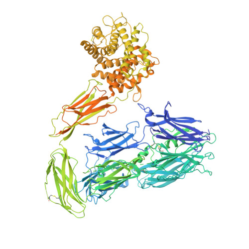
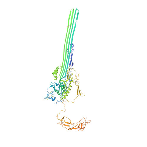
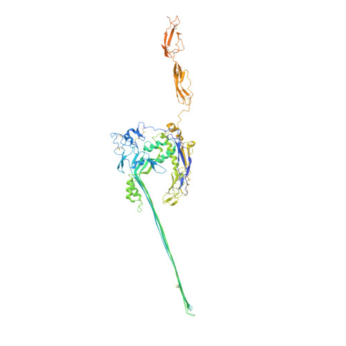
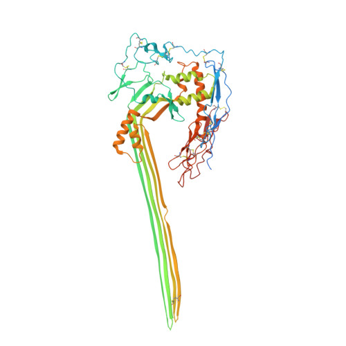
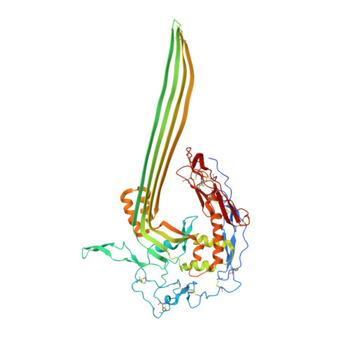
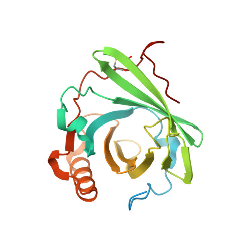
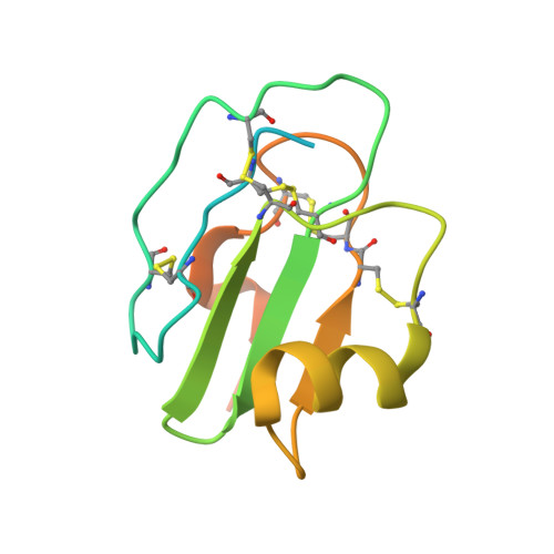
    CD59 is an abundant immuno-regulatory receptor that protects human cells from damage during complement activation. Here we show how the receptor binds complement proteins C8 and C9 at the membrane to prevent insertion and polymerization of membrane attack complex (MAC) pores. We present cryo-electron microscopy structures of two inhibited MAC precursors known as C5b8 and C5b9. We discover that in both complexes, CD59 binds the pore-forming β-hairpins of C8 to form an intermolecular β-sheet that prevents membrane perforation. While bound to C8, CD59 deflects the cascading C9 β-hairpins, rerouting their trajectory into the membrane. Preventing insertion of C9 restricts structural transitions of subsequent monomers and indirectly halts MAC polymerization. We combine our structural data with cellular assays and molecular dynamics simulations to explain how the membrane environment impacts the dual roles of CD59 in controlling pore formation of MAC, and as a target of bacterial virulence factors which hijack CD59 to lyse human cells.


  • Organizational Affiliation

    Department of Life Sciences, Sir Ernst Chain Building, Imperial College London, London, SW7 2AZ, United Kingdom.


Macromolecules
Find similar proteins by:  (by identity cutoff)  |  3D Structure
Entity ID: 1
MoleculeChains Sequence LengthOrganismDetailsImage
Complement C51,676Homo sapiensMutation(s): 0 
UniProt & NIH Common Fund Data Resources
Find proteins for P01031 (Homo sapiens)
Explore P01031 
Go to UniProtKB:  P01031
PHAROS:  P01031
GTEx:  ENSG00000106804 
Entity Groups  
Sequence Clusters30% Identity50% Identity70% Identity90% Identity95% Identity100% Identity
UniProt GroupP01031
Sequence Annotations
Expand
  • Reference Sequence
Find similar proteins by:  (by identity cutoff)  |  3D Structure
Entity ID: 2
MoleculeChains Sequence LengthOrganismDetailsImage
Complement component C6934Homo sapiensMutation(s): 0 
UniProt & NIH Common Fund Data Resources
Find proteins for P13671 (Homo sapiens)
Explore P13671 
Go to UniProtKB:  P13671
PHAROS:  P13671
GTEx:  ENSG00000039537 
Entity Groups  
Sequence Clusters30% Identity50% Identity70% Identity90% Identity95% Identity100% Identity
UniProt GroupP13671
Sequence Annotations
Expand
  • Reference Sequence
Find similar proteins by:  (by identity cutoff)  |  3D Structure
Entity ID: 3
MoleculeChains Sequence LengthOrganismDetailsImage
Complement component C7843Homo sapiensMutation(s): 0 
UniProt & NIH Common Fund Data Resources
Find proteins for P10643 (Homo sapiens)
Explore P10643 
Go to UniProtKB:  P10643
PHAROS:  P10643
GTEx:  ENSG00000112936 
Entity Groups  
Sequence Clusters30% Identity50% Identity70% Identity90% Identity95% Identity100% Identity
UniProt GroupP10643
Sequence Annotations
Expand
  • Reference Sequence
Find similar proteins by:  (by identity cutoff)  |  3D Structure
Entity ID: 4
MoleculeChains Sequence LengthOrganismDetailsImage
Complement component C8 beta chain591Homo sapiensMutation(s): 0 
UniProt & NIH Common Fund Data Resources
Find proteins for P07358 (Homo sapiens)
Explore P07358 
Go to UniProtKB:  P07358
PHAROS:  P07358
GTEx:  ENSG00000021852 
Entity Groups  
Sequence Clusters30% Identity50% Identity70% Identity90% Identity95% Identity100% Identity
UniProt GroupP07358
Sequence Annotations
Expand
  • Reference Sequence
Find similar proteins by:  (by identity cutoff)  |  3D Structure
Entity ID: 5
MoleculeChains Sequence LengthOrganismDetailsImage
Complement component C8 alpha chain584Homo sapiensMutation(s): 0 
UniProt & NIH Common Fund Data Resources
Find proteins for P07357 (Homo sapiens)
Explore P07357 
Go to UniProtKB:  P07357
PHAROS:  P07357
GTEx:  ENSG00000157131 
Entity Groups  
Sequence Clusters30% Identity50% Identity70% Identity90% Identity95% Identity100% Identity
UniProt GroupP07357
Sequence Annotations
Expand
  • Reference Sequence
Find similar proteins by:  (by identity cutoff)  |  3D Structure
Entity ID: 6
MoleculeChains Sequence LengthOrganismDetailsImage
Complement component C8 gamma chain202Homo sapiensMutation(s): 0 
UniProt & NIH Common Fund Data Resources
Find proteins for P07360 (Homo sapiens)
Explore P07360 
Go to UniProtKB:  P07360
PHAROS:  P07360
GTEx:  ENSG00000176919 
Entity Groups  
Sequence Clusters30% Identity50% Identity70% Identity90% Identity95% Identity100% Identity
UniProt GroupP07360
Sequence Annotations
Expand
  • Reference Sequence
Find similar proteins by:  (by identity cutoff)  |  3D Structure
Entity ID: 7
MoleculeChains Sequence LengthOrganismDetailsImage
CD59 glycoprotein120Escherichia coliMutation(s): 0 
Gene Names: CD59MIC11MIN1MIN2MIN3MSK21
UniProt & NIH Common Fund Data Resources
Find proteins for P13987 (Homo sapiens)
Explore P13987 
Go to UniProtKB:  P13987
PHAROS:  P13987
GTEx:  ENSG00000085063 
Entity Groups  
Sequence Clusters30% Identity50% Identity70% Identity90% Identity95% Identity100% Identity
UniProt GroupP13987
Sequence Annotations
Expand
  • Reference Sequence
Oligosaccharides

Help

Entity ID: 8
MoleculeChains Length2D Diagram Glycosylation3D Interactions
2-acetamido-2-deoxy-beta-D-glucopyranose-(1-4)-2-acetamido-2-deoxy-beta-D-glucopyranose
H, J
2N-Glycosylation
Glycosylation Resources
GlyTouCan:  G42666HT
GlyCosmos:  G42666HT
GlyGen:  G42666HT
Entity ID: 9
MoleculeChains Length2D Diagram Glycosylation3D Interactions
beta-D-mannopyranose-(1-4)-2-acetamido-2-deoxy-beta-D-glucopyranose
I
2N-Glycosylation
Glycosylation Resources
GlyTouCan:  G90333CG
GlyCosmos:  G90333CG
GlyGen:  G90333CG
Experimental Data & Validation

Experimental Data

  • Method: ELECTRON MICROSCOPY
  • Resolution: 3.00 Å
  • Aggregation State: PARTICLE 
  • Reconstruction Method: SINGLE PARTICLE 
EM Software:
TaskSoftware PackageVersion
RECONSTRUCTIONRELION3.1
MODEL REFINEMENTPHENIX1.20.1-4887
MODEL REFINEMENTISOLDE1.4
MODEL REFINEMENTCoot0.9.8.1

Structure Validation

View Full Validation Report


Entry History & Funding Information

Deposition Data


Funding OrganizationLocationGrant Number
European Research Council (ERC)European Union864751
Cancer Research UKUnited KingdomC24523/A26234

Revision History  (Full details and data files)

  • Version 1.0: 2023-02-22
    Type: Initial release
  • Version 1.1: 2023-03-01
    Changes: Database references