


Crystal structure of K449E variant of S-adenosyl-L-homocysteine hydrolase from Pseudomonas aeruginosa in complex with adenosine
Arning, A., Malecki, P., Wozniak, K., Brzezinski, K.To be published.
Experimental Data Snapshot
Starting Model: experimental
View more details
Entity ID: 1 | |||||
|---|---|---|---|---|---|
| Molecule | Chains | Sequence Length | Organism | Details | Image |
| Adenosylhomocysteinase | A, B [auth C] | 472 | Pseudomonas aeruginosa PAO1 | Mutation(s): 1 Gene Names: ahcY, PLES_04301 EC: 3.3.1.1 (PDB Primary Data), 3.13.2.1 (UniProt) | 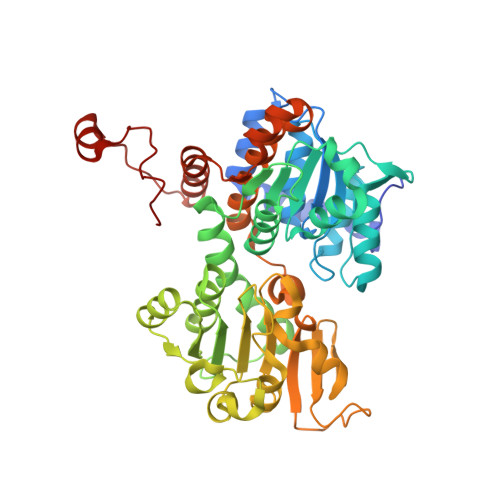 |
UniProt | |||||
Entity Groups | |||||
| Sequence Clusters | 30% Identity50% Identity70% Identity90% Identity95% Identity100% Identity | ||||
| UniProt Group | Q9I685 | ||||
Sequence AnnotationsExpand | |||||
Reference Sequence | |||||
| Ligands 4 Unique | |||||
|---|---|---|---|---|---|
| ID | Chains | Name / Formula / InChI Key | 2D Diagram | 3D Interactions | |
| NAD (Subject of Investigation/LOI) Download:Ideal Coordinates CCD File | C [auth A], G [auth C] | NICOTINAMIDE-ADENINE-DINUCLEOTIDE C21 H27 N7 O14 P2 BAWFJGJZGIEFAR-NNYOXOHSSA-N |  | ||
| ADN (Subject of Investigation/LOI) Download:Ideal Coordinates CCD File | D [auth A], H [auth C] | ADENOSINE C10 H13 N5 O4 OIRDTQYFTABQOQ-KQYNXXCUSA-N |  | ||
| PO4 Download:Ideal Coordinates CCD File | F [auth A], J [auth C] | PHOSPHATE ION O4 P NBIIXXVUZAFLBC-UHFFFAOYSA-K |  | ||
| K (Subject of Investigation/LOI) Download:Ideal Coordinates CCD File | E [auth A], I [auth C] | POTASSIUM ION K NPYPAHLBTDXSSS-UHFFFAOYSA-N |  | ||
| Length ( Å ) | Angle ( ˚ ) |
|---|---|
| a = 142.881 | α = 90 |
| b = 85.916 | β = 122.08 |
| c = 111.824 | γ = 90 |
| Software Name | Purpose |
|---|---|
| XDS | data reduction |
| XSCALE | data scaling |
| REFMAC | refinement |
| PDB_EXTRACT | data extraction |
| PHASER | phasing |
| Funding Organization | Location | Grant Number |
|---|---|---|
| Polish National Science Centre | Poland | SONATA BIS 2018/30/E/NZ1/00729 |


