Structural evolution of fibril polymorphs during amyloid assembly.
Wilkinson, M., Xu, Y., Thacker, D., Taylor, A.I.P., Fisher, D.G., Gallardo, R.U., Radford, S.E., Ranson, N.A.(2023) Cell 186: 5798-5811.e26
- PubMed: 38134875 Search on PubMed
- DOI: https://doi.org/10.1016/j.cell.2023.11.025
- Primary Citation Related Structures:
8AWT, 8AZ0, 8AZ1, 8AZ2, 8AZ3, 8AZ4, 8AZ5, 8AZ6, 8AZ7 - PubMed Abstract:
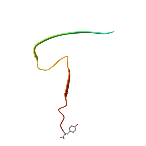
Cryoelectron microscopy (cryo-EM) has provided unprecedented insights into amyloid fibril structures, including those associated with disease. However, these structures represent the endpoints of long assembly processes, and their relationship to fibrils formed early in assembly is unknown. Consequently, whether different fibril architectures, with potentially different pathological properties, form during assembly remains unknown. Here, we used cryo-EM to determine structures of amyloid fibrils at different times during in vitro fibrillation of a disease-related variant of human islet amyloid polypeptide (IAPP-S20G). Strikingly, the fibrils formed in the lag, growth, and plateau phases have different structures, with new forms appearing and others disappearing as fibrillation proceeds. A time course with wild-type hIAPP also shows fibrils changing with time, suggesting that this is a general property of IAPP amyloid assembly. The observation of transiently populated fibril structures has implications for understanding amyloid assembly mechanisms with potential new insights into amyloid progression in disease.
- Astbury Centre for Structural Molecular Biology, School of Molecular & Cellular Biology, Faculty of Biological Sciences, University of Leeds, Leeds LS2 9JT, UK.
Organizational Affiliation: