8AY1 | pdb_00008ay1

Crystal structure of the C. elegans POFUT2 (CePoFUT2) triple mutant (R298K-R299K-A418C) in complex with the Rattus norvegicus TSR4 single mutant (E10C) from F-spondin


Experimental Data Snapshot

  • Method: X-RAY DIFFRACTION
  • Resolution: 2.13 Å
  • R-Value Free: 
    0.240 (Depositor), 0.242 (DCC) 
  • R-Value Work: 
    0.194 (Depositor), 0.201 (DCC) 
  • R-Value Observed: 
    0.195 (Depositor) 

Starting Model: experimental
View more details

wwPDB Validation 3D Report Full Report

Validation slider image for 8AY1

Ligand Structure Quality Assessment 


This is version 1.5 of the entry. See complete history

Literature

The Essential Role of Water Molecules in the Reaction Mechanism of Protein O-Fucosyltransferase 2.

Sanz-Martinez, I.Garcia-Garcia, A.Tejero, T.Hurtado-Guerrero, R.Merino, P.

(2022) Angew Chem Int Ed Engl 61: e202213610-e202213610

  • DOI: https://doi.org/10.1002/anie.202213610
  • Primary Citation Related Structures: 
    8AY1

  • PubMed Abstract: 

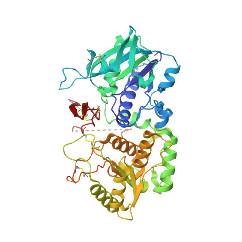
    Protein O-fucosyltransferase 2 (PoFUT2) is an inverting glycosyltransferase (GT) that fucosylates thrombospondin repeats (TSRs) from group 1 and 2. PoFUT2 recognizes a large and diverse number of TSRs through a dynamic network of water-mediated interactions. By X-ray structural studies of C. elegans PoFUT2 complexed to a TSR of group 2, we demonstrate that this GT recognizes similarly the 3D structure of TSRs from both groups 1 and 2. Its active site is highly exposed to the solvent, suggesting that water molecules might also play an essential role in the fucosylation mechanism. We applied QM/MM methods using human PoFUT2 as a model, and found that HsPoFUT2 follows a classical S N 2 reaction mechanism in which water molecules contribute to a great extent in facilitating the release of the leaving pyrophosphate unit, causing the H transfer from the acceptor nucleophile (Thr/Ser) to the catalytic base, which is the last event in the reaction. This demonstrates the importance of water molecules not only in recognition of the ligands but also in catalysis.


  • Organizational Affiliation
    • Instituto de Biocomputación y Física de Sistemas Complejos (BIFI)., Universidad de Zaragoza, 50018, Zaragoza, Spain.

Macromolecule Content 

  • Total Structure Weight: 109.01 kDa 
  • Atom Count: 7,237 
  • Modeled Residue Count: 830 
  • Deposited Residue Count: 924 
  • Unique protein chains: 1

Macromolecules

Find similar proteins by:|  3D Structure
Entity ID: 1
MoleculeChains  Sequence LengthOrganismDetailsImage
GDP-fucose protein O-fucosyltransferase 2,Spondin-1
A, B
462Caenorhabditis elegansMutation(s): 4 
Gene Names: pad-2K10G9.3Spon1rCG_39552
EC: 2.4.1.221
UniProt
Find proteins for P35446 (Rattus norvegicus)
Explore P35446 
Go to UniProtKB:  P35446
Find proteins for Q8WR51 (Caenorhabditis elegans)
Explore Q8WR51 
Go to UniProtKB:  Q8WR51
Entity Groups
Sequence Clusters30% Identity50% Identity70% Identity90% Identity95% Identity100% Identity
UniProt GroupsQ8WR51P35446
Glycosylation
Glycosylation Sites: 1Go to GlyGen: P35446-1
Sequence Annotations
Expand
Reference Sequence

Small Molecules

Ligands 4 Unique
IDChains Name / Formula / InChI Key2D Diagram3D Interactions
NAG

Query on NAG


Download:Ideal Coordinates CCD File
C [auth A],
P [auth B]
2-acetamido-2-deoxy-beta-D-glucopyranose
C8 H15 N O6
OVRNDRQMDRJTHS-FMDGEEDCSA-N
SO4

Query on SO4


Download:Ideal Coordinates CCD File
AA [auth B]
BA [auth B]
J [auth A]
K [auth A]
L [auth A]
AA [auth B],
BA [auth B],
J [auth A],
K [auth A],
L [auth A],
M [auth A],
N [auth A],
O [auth A],
W [auth B],
X [auth B],
Y [auth B],
Z [auth B]
SULFATE ION
O4 S
QAOWNCQODCNURD-UHFFFAOYSA-L
GOL

Query on GOL


Download:Ideal Coordinates CCD File
Q [auth B]GLYCEROL
C3 H8 O3
PEDCQBHIVMGVHV-UHFFFAOYSA-N
EDO

Query on EDO


Download:Ideal Coordinates CCD File
D [auth A]
E [auth A]
F [auth A]
G [auth A]
H [auth A]
D [auth A],
E [auth A],
F [auth A],
G [auth A],
H [auth A],
I [auth A],
R [auth B],
S [auth B],
T [auth B],
U [auth B],
V [auth B]
1,2-ETHANEDIOL
C2 H6 O2
LYCAIKOWRPUZTN-UHFFFAOYSA-N

Experimental Data & Validation

Experimental Data

  • Method: X-RAY DIFFRACTION
  • Resolution: 2.13 Å
  • R-Value Free:  0.240 (Depositor), 0.242 (DCC) 
  • R-Value Work:  0.194 (Depositor), 0.201 (DCC) 
  • R-Value Observed: 0.195 (Depositor) 
Space Group: P 63
Unit Cell:
Length ( Å )Angle ( ˚ )
a = 156.848α = 90
b = 156.848β = 90
c = 80.451γ = 120
Software Package:
Software NamePurpose
REFMACrefinement
XDSdata reduction
SCALAdata scaling
PHASERphasing

Structure Validation

View Full Validation Report


Ligand Structure Quality Assessment 


Entry History 

& Funding Information

Deposition Data


Funding OrganizationLocationGrant Number
Spanish Ministry of Science, Innovation, and UniversitiesSpain--

Revision History  (Full details and data files)

  • Version 1.0: 2022-10-26
    Type: Initial release
  • Version 1.1: 2022-11-02
    Changes: Database references
  • Version 1.2: 2022-11-23
    Changes: Database references
  • Version 1.3: 2022-11-30
    Changes: Database references
  • Version 1.4: 2024-01-31
    Changes: Data collection, Refinement description
  • Version 1.5: 2024-10-09
    Changes: Structure summary