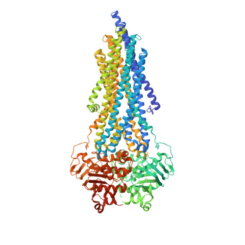
Tracing the substrate translocation mechanism in P-glycoprotein.
Gewering, T., Waghray, D., Parey, K., Jung, H., Tran, N.N.B., Zapata, J., Zhao, P., Chen, H., Januliene, D., Hummer, G., Urbatsch, I., Moeller, A., Zhang, Q.(2024) Elife 12
- PubMed: 38259172 Search on PubMedSearch on PubMed Central
- DOI: https://doi.org/10.7554/eLife.90174
- Primary Citation Related Structures:
7ZK4, 7ZK5, 7ZK6, 7ZK8, 7ZK9, 7ZKA, 7ZKB, 8AVY, 8PEE - PubMed Abstract:
P-glycoprotein (Pgp) is a prototypical ATP-binding cassette (ABC) transporter of great biological and clinical significance.Pgp confers cancer multidrug resistance and mediates the bioavailability and pharmacokinetics of many drugs (Juliano and Ling, 1976; Ueda et al., 1986; Sharom, 2011). Decades of structural and biochemical studies have provided insights into how Pgp binds diverse compounds (Loo and Clarke, 2000; Loo et al., 2009; Aller et al., 2009; Alam et al., 2019; Nosol et al., 2020; Chufan et al., 2015), but how they are translocated through the membrane has remained elusive. Here, we covalently attached a cyclic substrate to discrete sites of Pgp and determined multiple complex structures in inward- and outward-facing states by cryoEM. In conjunction with molecular dynamics simulations, our structures trace the substrate passage across the membrane and identify conformational changes in transmembrane helix 1 (TM1) as regulators of substrate transport. In mid-transport conformations, TM1 breaks at glycine 72. Mutation of this residue significantly impairs drug transport of Pgp in vivo, corroborating the importance of its regulatory role. Importantly, our data suggest that the cyclic substrate can exit Pgp without the requirement of a wide-open outward-facing conformation, diverting from the common efflux model for Pgp and other ABC exporters. The substrate transport mechanism of Pgp revealed here pinpoints critical targets for future drug discovery studies of this medically relevant system.
- Osnabrück University, Department of Biology/Chemistry, Structural Biology Section, Osnabrück, Germany.
Organizational Affiliation: