8AVQ | pdb_00008avq

AO75L in Complex with UDP-Xylose


Experimental Data Snapshot

  • Method: X-RAY DIFFRACTION
  • Resolution: 2.00 Å
  • R-Value Free: 
    0.227 (Depositor), 0.235 (DCC) 
  • R-Value Work: 
    0.196 (Depositor), 0.203 (DCC) 
  • R-Value Observed: 
    0.197 (Depositor) 

Starting Model: experimental
View more details

wwPDB Validation 3D Report Full Report

Validation slider image for 8AVQ

Ligand Structure Quality Assessment 


This is version 1.4 of the entry. See complete history

Literature

Unveiling the GT114 family: Structural characterization of A075L, a glycosyltransferase from Paramecium bursaria chlorella virus-1 (PBCV-1).

Laugieri, M.E.Speciale, I.Gimeno, A.Lin, S.Byers, B.W.Poveda, A.Nunez-Franco, R.Iturrioz, I.Moure, M.J.Jimenez-Oses, G.Russo-Krauss, I.Notaro, A.Van Etten, J.L.Lowary, T.L.Jimenez-Barbero, J.De Castro, C.Tonetti, M.Rojas, A.L.

(2024) Protein Sci 33: e5196-e5196

  • DOI: https://doi.org/10.1002/pro.5196
  • Primary Citation Related Structures: 
    8ASA, 8AVQ, 8Q8I

  • PubMed Abstract: 

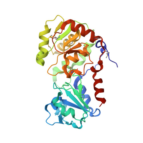
    Protein A075L is a β-xylosyltransferase that participates in producing the core of the N-glycans found in VP54, the major viral capsid protein of Paramecium bursaria chlorella virus-1 (PBCV-1). In this study, we present an X-ray crystallographic analysis of the apo form of A075L, along with its complexes with the sugar donor and with a trisaccharide acceptor. The protein structure shows a typical GT-B folding, with two Rossmann-like fold domains, in which the acceptor substrate binds to the N-terminal region, and the nucleotide-sugar donor binds to the C-terminal region. We propose that the catalytic mechanism follows a direct displacement S N 2-like reaction, where Asp73 serves as a catalytic base that deprotonates the incoming nucleophile of the acceptor, facilitating direct displacement of the UDP with the inversion of the anomeric configuration of the acceptor without metal ion dependence, while the interactions with side chains of Arg158 and Arg208 stabilize the developing negative charge. Using isothermal titration calorimetry, nuclear magnetic resonance spectroscopy, high-performance liquid chromatography, and molecular dynamics simulations, the catalytic activity and specificity of this enzyme have been unraveled.


  • Organizational Affiliation
    • DIMES - Biochemistry Division, University of Genoa, Genoa, Italy.

Macromolecule Content 

  • Total Structure Weight: 140.69 kDa 
  • Atom Count: 10,513 
  • Modeled Residue Count: 1,122 
  • Deposited Residue Count: 1,132 
  • Unique protein chains: 1

Macromolecules

Find similar proteins by:|  3D Structure
Entity ID: 1
MoleculeChains  Sequence LengthOrganismDetailsImage
AO75L
A, B, C, D
283Paramecium bursaria Chlorella virus 1Mutation(s): 0 
Gene Names: A075L
UniProt
Find proteins for Q89410 (Paramecium bursaria Chlorella virus 1)
Explore Q89410 
Go to UniProtKB:  Q89410
Entity Groups
Sequence Clusters30% Identity50% Identity70% Identity90% Identity95% Identity100% Identity
UniProt GroupQ89410
Sequence Annotations
Expand
Reference Sequence

Small Molecules

Ligands 6 Unique
IDChains Name / Formula / InChI Key2D Diagram3D Interactions
UDX
(Subject of Investigation/LOI)

Query on UDX


Download:Ideal Coordinates CCD File
H [auth A],
PB [auth D],
WA [auth C],
X [auth B]
URIDINE-5'-DIPHOSPHATE-XYLOPYRANOSE
C14 H22 N2 O16 P2
DQQDLYVHOTZLOR-OCIMBMBZSA-N
BCN

Query on BCN


Download:Ideal Coordinates CCD File
E [auth A],
MB [auth D],
RA [auth C],
V [auth B]
BICINE
C6 H13 N O4
FSVCELGFZIQNCK-UHFFFAOYSA-N
GOL

Query on GOL


Download:Ideal Coordinates CCD File
QA [auth B],
U [auth A],
W [auth B]
GLYCEROL
C3 H8 O3
PEDCQBHIVMGVHV-UHFFFAOYSA-N
EDO

Query on EDO


Download:Ideal Coordinates CCD File
DC [auth D]
EC [auth D]
F [auth A]
G [auth A]
LB [auth C]
DC [auth D],
EC [auth D],
F [auth A],
G [auth A],
LB [auth C],
NA [auth B],
NB [auth D],
OA [auth B],
OB [auth D],
PA [auth B],
S [auth A],
SA [auth C],
T [auth A],
TA [auth C],
UA [auth C],
VA [auth C]
1,2-ETHANEDIOL
C2 H6 O2
LYCAIKOWRPUZTN-UHFFFAOYSA-N
CA

Query on CA


Download:Ideal Coordinates CCD File
AA [auth B]
BA [auth B]
CA [auth B]
I [auth A]
J [auth A]
AA [auth B],
BA [auth B],
CA [auth B],
I [auth A],
J [auth A],
QB [auth D],
RB [auth D],
SB [auth D],
TB [auth D],
XA [auth C],
Y [auth B],
YA [auth C],
Z [auth B]
CALCIUM ION
Ca
BHPQYMZQTOCNFJ-UHFFFAOYSA-N
MG

Query on MG


Download:Ideal Coordinates CCD File
AB [auth C]
AC [auth D]
BB [auth C]
BC [auth D]
CB [auth C]
AB [auth C],
AC [auth D],
BB [auth C],
BC [auth D],
CB [auth C],
CC [auth D],
DA [auth B],
DB [auth C],
EA [auth B],
EB [auth C],
FA [auth B],
FB [auth C],
GA [auth B],
GB [auth C],
HA [auth B],
HB [auth C],
IA [auth B],
IB [auth C],
JA [auth B],
JB [auth C],
K [auth A],
KA [auth B],
KB [auth C],
L [auth A],
LA [auth B],
M [auth A],
MA [auth B],
N [auth A],
O [auth A],
P [auth A],
Q [auth A],
R [auth A],
UB [auth D],
VB [auth D],
WB [auth D],
XB [auth D],
YB [auth D],
ZA [auth C],
ZB [auth D]
MAGNESIUM ION
Mg
JLVVSXFLKOJNIY-UHFFFAOYSA-N
Modified Residues  1 Unique
IDChains TypeFormula2D DiagramParent
MSE
Query on MSE
A, B, C, D
L-PEPTIDE LINKINGC5 H11 N O2 SeMET

Experimental Data & Validation

Experimental Data

  • Method: X-RAY DIFFRACTION
  • Resolution: 2.00 Å
  • R-Value Free:  0.227 (Depositor), 0.235 (DCC) 
  • R-Value Work:  0.196 (Depositor), 0.203 (DCC) 
  • R-Value Observed: 0.197 (Depositor) 
Space Group: C 1 2 1
Unit Cell:
Length ( Å )Angle ( ˚ )
a = 129.285α = 90
b = 130.427β = 118.01
c = 87.143γ = 90
Software Package:
Software NamePurpose
XDSdata reduction
XSCALEdata scaling
REFMACrefinement
PDB_EXTRACTdata extraction
MOLREPphasing

Structure Validation

View Full Validation Report


Ligand Structure Quality Assessment 


Entry History 

& Funding Information

Deposition Data


Funding OrganizationLocationGrant Number
Spanish Ministry of Science, Innovation, and UniversitiesSpainSEV20160644

Revision History  (Full details and data files)

  • Version 1.0: 2023-09-06
    Type: Initial release
  • Version 1.1: 2023-11-15
    Changes: Data collection
  • Version 1.2: 2024-10-16
    Changes: Structure summary
  • Version 1.3: 2024-11-27
    Changes: Database references
  • Version 1.4: 2026-03-04
    Changes: Refinement description