8ARR | pdb_00008arr

Small molecular stabilizer for ERalpha and 14-3-3 (1076394)


Experimental Data Snapshot

  • Method: X-RAY DIFFRACTION
  • Resolution: 1.35 Å
  • R-Value Free: 
    0.199 (Depositor), 0.199 (DCC) 
  • R-Value Work: 
    0.179 (Depositor), 0.178 (DCC) 
  • R-Value Observed: 
    0.180 (Depositor) 

Starting Model: experimental
View more details

wwPDB Validation 3D Report Full Report

Validation slider image for 8ARR

Ligand Structure Quality Assessment 


This is version 1.3 of the entry. See complete history

Literature

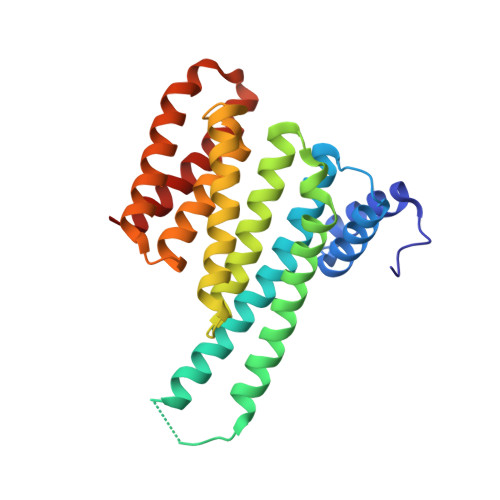
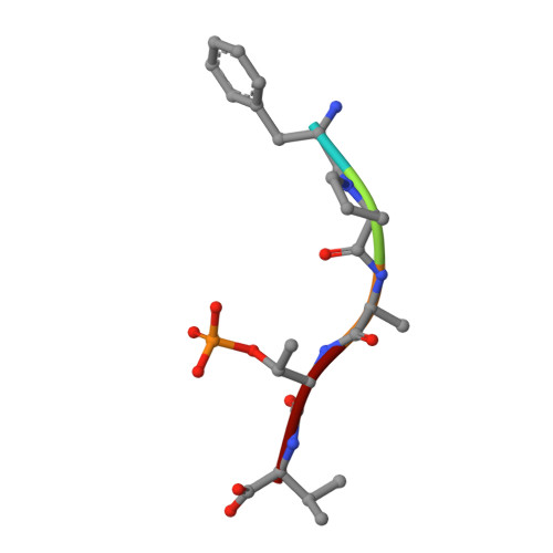
Structure-Based Optimization of Covalent, Small-Molecule Stabilizers of the 14-3-3 sigma /ER alpha Protein-Protein Interaction from Nonselective Fragments.

Konstantinidou, M.Visser, E.J.Vandenboorn, E.Chen, S.Jaishankar, P.Overmans, M.Dutta, S.Neitz, R.J.Renslo, A.R.Ottmann, C.Brunsveld, L.Arkin, M.R.

(2023) J Am Chem Soc 145: 20328-20343

  • DOI: https://doi.org/10.1021/jacs.3c05161
  • Primary Citation Related Structures: 
    8AI0, 8ALR, 8ALT, 8ALV, 8ALW, 8AM7, 8ANF, 8AOY, 8APS, 8AQ1, 8AQC, 8AQE, 8AQZ, 8AR4, 8AR5, 8ARG, 8ARO, 8ARQ, 8ARR, 8ARW, 8ARX, 8ARY, 8ARZ, 8AS1, 8AT9, 8ATP, 8ATR, 8ATS, 8AU2, 8AUS, 8AUY, 8AV3, 8AV4, 8AV7, 8AV8, 8AWG, 8AXE, 8AXU, 8AZE, 8B39

  • PubMed Abstract: 

    The stabilization of protein-protein interactions (PPIs) has emerged as a promising strategy in chemical biology and drug discovery. The identification of suitable starting points for stabilizing native PPIs and their subsequent elaboration into selective and potent molecular glues lacks structure-guided optimization strategies. We have previously identified a disulfide fragment that stabilized the hub protein 14-3-3σ bound to several of its clients, including ERα and C-RAF. Here, we show the structure-based optimization of the nonselective fragment toward selective and highly potent small-molecule stabilizers of the 14-3-3σ/ERα complex. The more elaborated molecular glues, for example, show no stabilization of 14-3-3σ/C-RAF up to 150 μM compound. Orthogonal biophysical assays, including mass spectrometry and fluorescence anisotropy, were used to establish structure-activity relationships. The binding modes of 37 compounds were elucidated with X-ray crystallography, which further assisted the concomitant structure-guided optimization. By targeting specific amino acids in the 14-3-3σ/ERα interface and locking the conformation with a spirocycle, the optimized covalent stabilizer 181 achieved potency, cooperativity, and selectivity similar to the natural product Fusicoccin-A. This case study showcases the value of addressing the structure, kinetics, and cooperativity for molecular glue development.


  • Organizational Affiliation
    • Department of Pharmaceutical Chemistry and Small Molecule Discovery Center (SMDC), University of California, San Francisco, California 94143, United States.

Macromolecule Content 

  • Total Structure Weight: 27.65 kDa 
  • Atom Count: 2,175 
  • Modeled Residue Count: 238 
  • Deposited Residue Count: 241 
  • Unique protein chains: 2

Macromolecules

Find similar proteins by:|  3D Structure
Entity ID: 1
MoleculeChains  Sequence LengthOrganismDetailsImage
14-3-3 protein sigma236Homo sapiensMutation(s): 0 
Gene Names: SFNHME1
UniProt & NIH Common Fund Data Resources
Find proteins for P31947 (Homo sapiens)
Explore P31947 
Go to UniProtKB:  P31947
PHAROS:  P31947
GTEx:  ENSG00000175793 
Entity Groups
Sequence Clusters30% Identity50% Identity70% Identity90% Identity95% Identity100% Identity
UniProt GroupP31947
Sequence Annotations
Expand
Reference Sequence
Find similar proteins by:  Sequence   |   3D Structure  
Entity ID: 2
MoleculeChains  Sequence LengthOrganismDetailsImage
Estrogen receptor5Homo sapiensMutation(s): 0 
UniProt & NIH Common Fund Data Resources
Find proteins for P03372 (Homo sapiens)
Explore P03372 
Go to UniProtKB:  P03372
PHAROS:  P03372
GTEx:  ENSG00000091831 
Entity Groups
UniProt GroupP03372
Sequence Annotations
Expand
Reference Sequence

Small Molecules

Modified Residues  1 Unique
IDChains TypeFormula2D DiagramParent
TPO
Query on TPO
B
L-PEPTIDE LINKINGC4 H10 N O6 PTHR

Experimental Data & Validation

Experimental Data

  • Method: X-RAY DIFFRACTION
  • Resolution: 1.35 Å
  • R-Value Free:  0.199 (Depositor), 0.199 (DCC) 
  • R-Value Work:  0.179 (Depositor), 0.178 (DCC) 
  • R-Value Observed: 0.180 (Depositor) 
Space Group: C 2 2 21
Unit Cell:
Length ( Å )Angle ( ˚ )
a = 81.577α = 90
b = 111.964β = 90
c = 62.269γ = 90
Software Package:
Software NamePurpose
REFMACrefinement
DIALSdata reduction
Aimlessdata scaling
MOLREPphasing

Structure Validation

View Full Validation Report


Ligand Structure Quality Assessment 


Entry History 

& Funding Information

Deposition Data


Funding OrganizationLocationGrant Number
Netherlands Organisation for Scientific Research (NWO)Netherlands--

Revision History  (Full details and data files)

  • Version 1.0: 2023-09-20
    Type: Initial release
  • Version 1.1: 2023-10-04
    Changes: Database references
  • Version 1.2: 2024-11-06
    Changes: Structure summary
  • Version 1.3: 2026-03-04
    Changes: Refinement description