


Crystal structure of the Q65N mutant of S-adenosyl-L-homocysteine hydrolase from Pseudomonas aeruginosa crystallized in the presence of K+ cations
Drozdzal, P., Wozniak, K., Malecki, P., Gawel, M., Komorowska, M., Brzezinski, K.To be published.
Experimental Data Snapshot
Starting Model: experimental
View more details
Entity ID: 1 | |||||
|---|---|---|---|---|---|
| Molecule | Chains | Sequence Length | Organism | Details | Image |
| Adenosylhomocysteinase | 472 | Pseudomonas aeruginosa PAO1 | Mutation(s): 1 Gene Names: ahcY, sahH, PA0432 EC: 3.3.1.1 (PDB Primary Data), 3.13.2.1 (UniProt) | 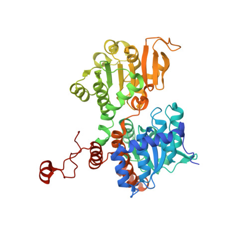 | |
UniProt | |||||
Entity Groups | |||||
| Sequence Clusters | 30% Identity50% Identity70% Identity90% Identity95% Identity100% Identity | ||||
| UniProt Group | Q9I685 | ||||
Sequence AnnotationsExpand | |||||
Reference Sequence | |||||
| Ligands 5 Unique | |||||
|---|---|---|---|---|---|
| ID | Chains | Name / Formula / InChI Key | 2D Diagram | 3D Interactions | |
| NAD (Subject of Investigation/LOI) Download:Ideal Coordinates CCD File | AB [auth III] CA [auth DDD] I [auth AAA] MA [auth FFF] N [auth BBB] | NICOTINAMIDE-ADENINE-DINUCLEOTIDE C21 H27 N7 O14 P2 BAWFJGJZGIEFAR-NNYOXOHSSA-N |  | ||
| PEG Download:Ideal Coordinates CCD File | AA [auth CCC], BA [auth CCC] | DI(HYDROXYETHYL)ETHER C4 H10 O3 MTHSVFCYNBDYFN-UHFFFAOYSA-N |  | ||
| PO4 Download:Ideal Coordinates CCD File | GA [auth DDD] HA [auth DDD] IA [auth DDD] JA [auth FFF] K [auth AAA] | PHOSPHATE ION O4 P NBIIXXVUZAFLBC-UHFFFAOYSA-K |  | ||
| GOL Download:Ideal Coordinates CCD File | BB [auth III] CB [auth III] DA [auth DDD] EA [auth DDD] FA [auth DDD] | GLYCEROL C3 H8 O3 PEDCQBHIVMGVHV-UHFFFAOYSA-N |  | ||
| K (Subject of Investigation/LOI) Download:Ideal Coordinates CCD File | V [auth BBB] | POTASSIUM ION K NPYPAHLBTDXSSS-UHFFFAOYSA-N |  | ||
| Length ( Å ) | Angle ( ˚ ) |
|---|---|
| a = 111.481 | α = 90 |
| b = 211.877 | β = 105.325 |
| c = 111.351 | γ = 90 |
| Software Name | Purpose |
|---|---|
| REFMAC | refinement |
| XDS | data reduction |
| XSCALE | data scaling |
| PHASER | phasing |
| Funding Organization | Location | Grant Number |
|---|---|---|
| Polish National Science Centre | Poland | OPUS 2013/09/B/NZ1/01880 |


