8AF0

Crystal structure of human angiogenin and RNA duplex


Experimental Data Snapshot

  • Method: X-RAY DIFFRACTION
  • Resolution: 2.43 Å
  • R-Value Free: 0.268 
  • R-Value Work: 0.205 
  • R-Value Observed: 0.208 

wwPDB Validation   3D Report Full Report


This is version 1.1 of the entry. See complete history


Literature

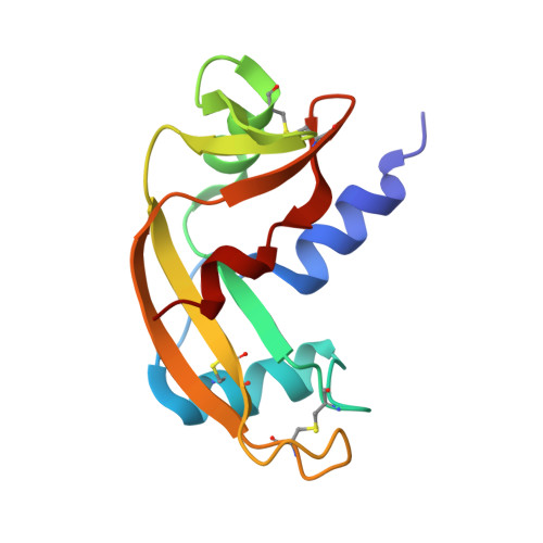
Structure of angiogenin dimer bound to double-stranded RNA.

Sievers, K.Ficner, R.

(2022) Acta Crystallogr F Struct Biol Commun 78: 330-337

  • DOI: https://doi.org/10.1107/S2053230X22008317
  • Primary Citation of Related Structures:  
    8AF0

  • PubMed Abstract: 

    Angiogenin is an unusual member of the RNase A family and is of great interest in multiple pathological contexts. Although it has been assigned various regulatory roles, its core catalytic function is that of an RNA endonuclease. However, its catalytic efficiency is comparatively low and this has been linked to a unique C-terminal helix which partially blocks its RNA-binding site. Assuming that binding to its RNA substrate could trigger a conformational rearrangement, much speculation has arisen on the topic of the interaction of angiogenin with RNA. To date, no structural data on angiogenin-RNA interactions have been available. Here, the structure of angiogenin bound to a double-stranded RNA duplex is reported. The RNA does not reach the active site of angiogenin and no structural arrangement of the C-terminal domain is observed. However, angiogenin forms a previously unobserved crystallographic dimer that makes several backbone interactions with the major and minor grooves of the RNA double helix.


  • Organizational Affiliation

    Department for Molecular Structural Biology, Georg-August-Universität Göttingen, Justus-von-Liebig Weg 11, 37077 Göttingen, Germany.


Macromolecules

Find similar proteins by:  (by identity cutoff)  |  3D Structure
Entity ID: 2
MoleculeChains Sequence LengthOrganismDetailsImage
AngiogeninC [auth A],
D [auth B]
123Homo sapiensMutation(s): 1 
Gene Names: ANGRNASE5
EC: 3.1.27
UniProt & NIH Common Fund Data Resources
Find proteins for P03950 (Homo sapiens)
Explore P03950 
Go to UniProtKB:  P03950
PHAROS:  P03950
GTEx:  ENSG00000214274 
Entity Groups  
Sequence Clusters30% Identity50% Identity70% Identity90% Identity95% Identity100% Identity
UniProt GroupP03950
Sequence Annotations
Expand
  • Reference Sequence

Find similar nucleic acids by:  Sequence   |   3D Structure  

Entity ID: 1
MoleculeChains LengthOrganismImage
RNA (5'-R(*GP*CP*CP*CP*GP*CP*CP*UP*GP*UP*CP*AP*CP*GP*CP*GP*GP*GP*C)-3')A [auth C],
B [auth D]
19Homo sapiens
Sequence Annotations
Expand
  • Reference Sequence
Experimental Data & Validation

Experimental Data

  • Method: X-RAY DIFFRACTION
  • Resolution: 2.43 Å
  • R-Value Free: 0.268 
  • R-Value Work: 0.205 
  • R-Value Observed: 0.208 
  • Space Group: P 21 21 21
Unit Cell:
Length ( Å )Angle ( ˚ )
a = 50.38α = 90
b = 66.66β = 90
c = 101.4γ = 90
Software Package:
Software NamePurpose
PHASERphasing
REFMACrefinement
PHENIXrefinement
XSCALEdata scaling
XDSdata reduction

Structure Validation

View Full Validation Report


Entry History & Funding Information

Deposition Data


Funding OrganizationLocationGrant Number
German Research Foundation (DFG)Germany--

Revision History  (Full details and data files)

  • Version 1.0: 2022-09-14
    Type: Initial release
  • Version 1.1: 2024-01-31
    Changes: Data collection, Refinement description