8AAV

Human heavy chain ferritin with introduced Cys residues modified with C10 ligand


Experimental Data Snapshot

  • Method: X-RAY DIFFRACTION
  • Resolution: 2.00 Å
  • R-Value Free: 0.177 
  • R-Value Work: 0.144 
  • R-Value Observed: 0.145 

wwPDB Validation   3D Report Full Report


Ligand Structure Quality Assessment 


This is version 1.0 of the entry. See complete history


Literature

Assembly of chemically modified protein nanocages into 3D materials for the adsorption of uremic toxins.

Bohler, H.Orth-Alampour, S.Baaten, C.Riedner, M.Jankowski, J.Beck, T.

(2022) J Mater Chem B 11: 55-60

  • DOI: https://doi.org/10.1039/d2tb02386e
  • Primary Citation of Related Structures:  
    8AAV

  • PubMed Abstract: 

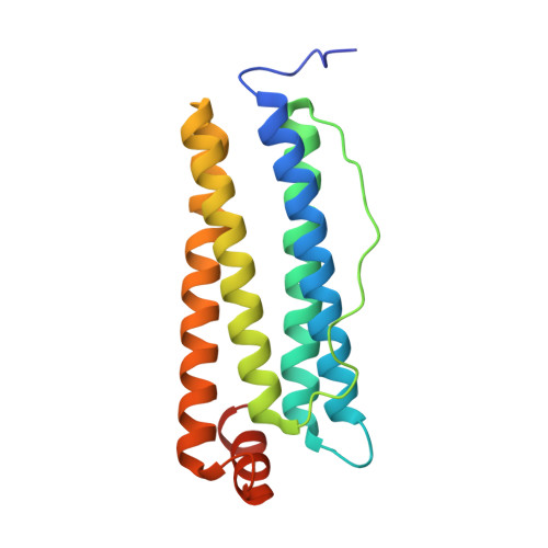
    Hemodialysis fails to remove protein-bound uremic toxins that are attributed with high cardiovascular risk. Application of adsorption materials is a viable strategy, but suitable biocompatible adsorbents are still not available. Here, we demonstrate that adsorbents based on the bottom-up assembly of the intrinsically biocompatible protein cage ferritin are applicable for toxin adsorption. Due to the size-exclusion effect of its pores, only small molecules such as uremic toxins can enter the protein cage. Protein redesign techniques that target selectively the inner surface were used to introduce anchor sites for chemical modification. Porous crystalline adsorbents were fabricated by bottom-up assembly of the protein cage. Linkage of up to 96 phenylic or aliphatic molecules per container was verified by ESI-MS. Materials based on unmodified ferritin cages can already adsorb the uremic toxins. The adsorption capacity could be increased by about 50% through functionalization with hydrophobic molecules reaching 458 μg g -1 for indoxyl sulfate. The biohybrid materials show no contamination with endotoxins and do not activate blood platelets. These findings demonstrate the great potential of protein-based adsorbents for the clearance of uremic toxins: modifications enhance toxin adsorption without diminishing the biocompatibility of the final protein-based material.


  • Organizational Affiliation

    Universität Hamburg, Department of Chemistry, Institute of Physical Chemistry, Grindelallee 117, Hamburg 20146, Germany. tobias.beck@chemie.uni-hamburg.de.


Macromolecules
Find similar proteins by:  (by identity cutoff)  |  3D Structure
Entity ID: 1
MoleculeChains Sequence LengthOrganismDetailsImage
Ferritin heavy chain, N-terminally processed
A, B, C, D, E
A, B, C, D, E, F, G, H
182Homo sapiensMutation(s): 0 
Gene Names: FTH1FTHFTHL6OK/SW-cl.84PIG15
UniProt & NIH Common Fund Data Resources
Find proteins for P02794 (Homo sapiens)
Explore P02794 
Go to UniProtKB:  P02794
PHAROS:  P02794
GTEx:  ENSG00000167996 
Entity Groups  
Sequence Clusters30% Identity50% Identity70% Identity90% Identity95% Identity100% Identity
UniProt GroupP02794
Sequence Annotations
Expand
  • Reference Sequence
Small Molecules
Ligands 3 Unique
IDChains Name / Formula / InChI Key2D Diagram3D Interactions
O3K
Query on O3K

Download Ideal Coordinates CCD File 
BA [auth E]
CA [auth E]
GA [auth F]
HA [auth F]
I [auth A]
BA [auth E],
CA [auth E],
GA [auth F],
HA [auth F],
I [auth A],
J [auth A],
MA [auth G],
N [auth B],
NA [auth G],
O [auth B],
RA [auth H],
SA [auth H],
T [auth C],
U [auth C],
Y [auth D],
Z [auth D]
2-bromanyl-N-decyl-ethanamide
C12 H24 Br N O
RZTGBAJAVREPOB-UHFFFAOYSA-N
FE
Query on FE

Download Ideal Coordinates CCD File 
AA [auth D]
DA [auth E]
IA [auth F]
K [auth A]
OA [auth G]
AA [auth D],
DA [auth E],
IA [auth F],
K [auth A],
OA [auth G],
P [auth B],
TA [auth H],
V [auth C]
FE (III) ION
Fe
VTLYFUHAOXGGBS-UHFFFAOYSA-N
MG
Query on MG

Download Ideal Coordinates CCD File 
EA [auth E]
FA [auth E]
JA [auth F]
KA [auth F]
L [auth A]
EA [auth E],
FA [auth E],
JA [auth F],
KA [auth F],
L [auth A],
LA [auth F],
M [auth A],
PA [auth G],
Q [auth B],
QA [auth G],
R [auth B],
S [auth B],
UA [auth H],
W [auth C],
X [auth C]
MAGNESIUM ION
Mg
JLVVSXFLKOJNIY-UHFFFAOYSA-N
Experimental Data & Validation

Experimental Data

  • Method: X-RAY DIFFRACTION
  • Resolution: 2.00 Å
  • R-Value Free: 0.177 
  • R-Value Work: 0.144 
  • R-Value Observed: 0.145 
  • Space Group: P 2 3
Unit Cell:
Length ( Å )Angle ( ˚ )
a = 181.44α = 90
b = 181.44β = 90
c = 181.44γ = 90
Software Package:
Software NamePurpose
Aimlessdata scaling
REFMACrefinement
PDB_EXTRACTdata extraction
XDSdata reduction
PHASERphasing

Structure Validation

View Full Validation Report


Ligand Structure Quality Assessment 


Entry History & Funding Information

Deposition Data


Funding OrganizationLocationGrant Number
German Research Foundation (DFG)Germany401323995
German Research Foundation (DFG)Germany413227073

Revision History  (Full details and data files)

  • Version 1.0: 2024-03-20
    Type: Initial release