


Crystal structure of CYP142 from Mycobacterium tuberculosis in complex with cholestenone
Snee, M., Amadi, C.To be published.
Experimental Data Snapshot
Starting Model: experimental
View more details
Entity ID: 1 | |||||
|---|---|---|---|---|---|
| Molecule | Chains | Sequence Length | Organism | Details | Image |
| Steroid C26-monooxygenase | A [auth B], B [auth E] | 418 | Mycobacterium tuberculosis H37Rv | Mutation(s): 0 Gene Names: cyp142, cyp142A1, Rv3518c, MTV023.25c EC: 1.14.15.28 | 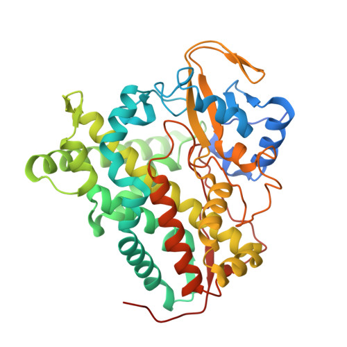 |
UniProt | |||||
Entity Groups | |||||
| Sequence Clusters | 30% Identity50% Identity70% Identity90% Identity95% Identity100% Identity | ||||
| UniProt Group | P9WPL5 | ||||
Sequence AnnotationsExpand | |||||
Reference Sequence | |||||
| Ligands 2 Unique | |||||
|---|---|---|---|---|---|
| ID | Chains | Name / Formula / InChI Key | 2D Diagram | 3D Interactions | |
| HEM Download:Ideal Coordinates CCD File | D [auth B], F [auth E] | PROTOPORPHYRIN IX CONTAINING FE C34 H32 Fe N4 O4 KABFMIBPWCXCRK-RGGAHWMASA-L |  | ||
| K2B (Subject of Investigation/LOI) Download:Ideal Coordinates CCD File | C [auth B], E | (8ALPHA,9BETA)-CHOLEST-4-EN-3-ONE C27 H44 O NYOXRYYXRWJDKP-GYKMGIIDSA-N |  | ||
| Length ( Å ) | Angle ( ˚ ) |
|---|---|
| a = 65.81 | α = 90 |
| b = 128.13 | β = 90 |
| c = 146.6 | γ = 90 |
| Software Name | Purpose |
|---|---|
| PHENIX | refinement |
| DIALS | data reduction |
| Aimless | data scaling |
| PHASER | phasing |
| Funding Organization | Location | Grant Number |
|---|---|---|
| Biotechnology and Biological Sciences Research Council (BBSRC) | United Kingdom | -- |


