8A4Y | pdb_00008a4y

SARS-CoV-2 non-structural protein-1 (nsp1) in complex with N-(2,3-dihydro-1H-inden-5-yl)acetamide


Experimental Data Snapshot

  • Method: X-RAY DIFFRACTION
  • Resolution: 1.10 Å
  • R-Value Free: 
    0.166 (Depositor), 0.156 (DCC) 
  • R-Value Work: 
    0.147 (Depositor), 0.148 (DCC) 
  • R-Value Observed: 
    0.148 (Depositor) 

Starting Model: experimental
View more details

wwPDB Validation 3D Report Full Report

Validation slider image for 8A4Y

Ligand Structure Quality Assessment 


This is version 1.2 of the entry. See complete history

Literature

Revealing druggable cryptic pockets in the Nsp1 of SARS-CoV-2 and other beta-coronaviruses by simulations and crystallography.

Borsatto, A.Akkad, O.Galdadas, I.Ma, S.Damfo, S.Haider, S.Kozielski, F.Estarellas, C.Gervasio, F.L.

(2022) Elife 11

  • DOI: https://doi.org/10.7554/eLife.81167
  • Primary Citation Related Structures: 
    8A4Y

  • PubMed Abstract: 

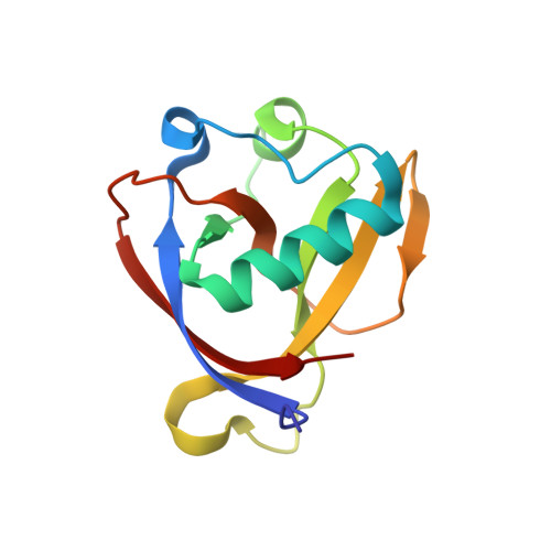
    Non-structural protein 1 (Nsp1) is a main pathogenicity factor of α - and β - coronaviruses. Nsp1 of severe acute respiratory syndrome coronavirus 2 (SARS-CoV-2) suppresses the host gene expression by sterically blocking 40S host ribosomal subunits and promoting host mRNA degradation. This mechanism leads to the downregulation of the translation-mediated innate immune response in host cells, ultimately mediating the observed immune evasion capabilities of SARS-CoV-2. Here, by combining extensive molecular dynamics simulations, fragment screening and crystallography, we reveal druggable pockets in Nsp1. Structural and computational solvent mapping analyses indicate the partial crypticity of these newly discovered and druggable binding sites. The results of fragment-based screening via X-ray crystallography confirm the druggability of the major pocket of Nsp1. Finally, we show how the targeting of this pocket could disrupt the Nsp1-mRNA complex and open a novel avenue to design new inhibitors for other Nsp1s present in homologous β - coronaviruses.


  • Organizational Affiliation
    • School of Pharmaceutical Sciences, University of Geneva, Geneva, Switzerland.

Macromolecule Content 

  • Total Structure Weight: 13.28 kDa 
  • Atom Count: 1,082 
  • Modeled Residue Count: 118 
  • Deposited Residue Count: 118 
  • Unique protein chains: 1

Macromolecules

Find similar proteins by:|  3D Structure
Entity ID: 1
MoleculeChains  Sequence LengthOrganismDetailsImage
Host translation inhibitor nsp1118Severe acute respiratory syndrome coronavirus 2Mutation(s): 0 
Gene Names: rep1a-1b
UniProt
Find proteins for P0DTD1 (Severe acute respiratory syndrome coronavirus 2)
Explore P0DTD1 
Go to UniProtKB:  P0DTD1
Entity Groups
Sequence Clusters30% Identity50% Identity70% Identity90% Identity95% Identity100% Identity
UniProt GroupP0DTD1
Sequence Annotations
Expand
Reference Sequence

Small Molecules

Ligands 1 Unique
IDChains Name / Formula / InChI Key2D Diagram3D Interactions
QO6
(Subject of Investigation/LOI)

Query on QO6


Download:Ideal Coordinates CCD File
B [auth A]N-(2,3-dihydro-1H-inden-5-yl)acetamide
C11 H13 N O
WOYOJAPRKMBKEU-UHFFFAOYSA-N

Experimental Data & Validation

Experimental Data

  • Method: X-RAY DIFFRACTION
  • Resolution: 1.10 Å
  • R-Value Free:  0.166 (Depositor), 0.156 (DCC) 
  • R-Value Work:  0.147 (Depositor), 0.148 (DCC) 
  • R-Value Observed: 0.148 (Depositor) 
Space Group: P 43 21 2
Unit Cell:
Length ( Å )Angle ( ˚ )
a = 36.688α = 90
b = 36.688β = 90
c = 140.928γ = 90
Software Package:
Software NamePurpose
PHENIXrefinement
PDB_EXTRACTdata extraction
autoPROCdata reduction
autoPROCdata scaling
PHENIXphasing

Structure Validation

View Full Validation Report


Ligand Structure Quality Assessment 


Entry History 

& Funding Information

Deposition Data


Funding OrganizationLocationGrant Number
Not funded--

Revision History  (Full details and data files)

  • Version 1.0: 2022-11-23
    Type: Initial release
  • Version 1.1: 2022-11-30
    Changes: Database references
  • Version 1.2: 2024-01-31
    Changes: Data collection, Refinement description