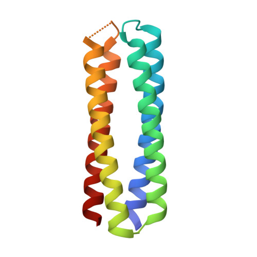
From peptides to proteins: coiled-coil tetramers to single-chain 4-helix bundles.
Naudin, E.A., Albanese, K.I., Smith, A.J., Mylemans, B., Baker, E.G., Weiner, O.D., Andrews, D.M., Tigue, N., Savery, N.J., Woolfson, D.N.(2022) Chem Sci 13: 11330-11340
- PubMed: 36320580 Search on PubMedSearch on PubMed Central
- DOI: https://doi.org/10.1039/d2sc04479j
- Primary Citation Related Structures:
8A3G, 8A3I, 8A3J, 8A3K - PubMed Abstract:
The design of completely synthetic proteins from first principles- de novo protein design-is challenging. This is because, despite recent advances in computational protein-structure prediction and design, we do not understand fully the sequence-to-structure relationships for protein folding, assembly, and stabilization. Antiparallel 4-helix bundles are amongst the most studied scaffolds for de novo protein design. We set out to re-examine this target, and to determine clear sequence-to-structure relationships, or design rules, for the structure. Our aim was to determine a common and robust sequence background for designing multiple de novo 4-helix bundles. In turn, this could be used in chemical and synthetic biology to direct protein-protein interactions and as scaffolds for functional protein design. Our approach starts by analyzing known antiparallel 4-helix coiled-coil structures to deduce design rules. In terms of the heptad repeat, abcdefg - i.e. , the sequence signature of many helical bundles-the key features that we identify are: a = Leu, d = Ile, e = Ala, g = Gln, and the use of complementary charged residues at b and c. Next, we implement these rules in the rational design of synthetic peptides to form antiparallel homo- and heterotetramers. Finally, we use the sequence of the homotetramer to derive in one step a single-chain 4-helix-bundle protein for recombinant production in E. coli . All of the assembled designs are confirmed in aqueous solution using biophysical methods, and ultimately by determining high-resolution X-ray crystal structures. Our route from peptides to proteins provides an understanding of the role of each residue in each design.
- School of Chemistry, University of Bristol Cantock's Close Bristol BS8 1TS UK d.n.woolfson@bristol.ac.uk.
Organizational Affiliation: