8VIS | pdb_00008vis

Human TMPRSS11D complexed with a disulfide-linked autoinhibitory DDDDK peptide


Experimental Data Snapshot

  • Method: X-RAY DIFFRACTION
  • Resolution: 1.59 Å
  • R-Value Free: 
    0.187 (Depositor), 0.195 (DCC) 
  • R-Value Work: 
    0.155 (Depositor), 0.168 (DCC) 
  • R-Value Observed: 
    0.155 (Depositor) 

Starting Model: experimental
View more details

wwPDB Validation 3D Report Full Report

Validation slider image for 8VIS

This is version 1.2 of the entry. See complete history

Literature

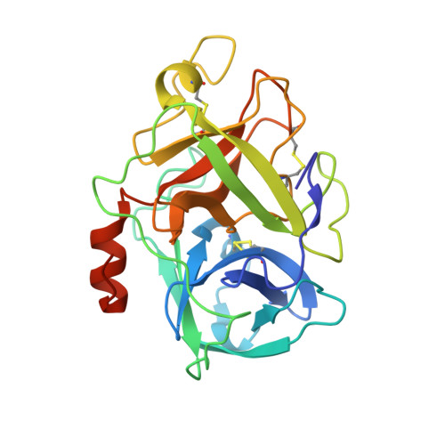
Structural basis of TMPRSS11D specificity and autocleavage activation.

Fraser, B.J.Wilson, R.P.Ferkova, S.Ilyassov, O.Lac, J.Dong, A.Li, Y.Y.Seitova, A.Li, Y.Hejazi, Z.Kenney, T.M.G.Penn, L.Z.Edwards, A.Leduc, R.Boudreault, P.L.Morin, G.B.Benard, F.Arrowsmith, C.H.

(2025) Nat Commun 16: 4351-4351

  • DOI: https://doi.org/10.1038/s41467-025-59677-3
  • Primary Citation Related Structures: 
    8VIS, 9DPF

  • PubMed Abstract: 

    Transmembrane Protease, Serine-2 (TMPRSS2) and TMPRSS11D are human proteases that enable SARS-CoV-2 and Influenza A/B virus infections, but their biochemical mechanisms for facilitating viral cell entry remain unclear. We show these proteases spontaneously and efficiently cleave their own zymogen activation motifs, activating their broader protease activity on cellular substrates. We determine TMPRSS11D co-crystal structures with a native and an engineered activation motif, revealing insights into its autocleavage activation and distinct substrate binding cleft features. Leveraging this structural data, we develop nanomolar potency peptidomimetic inhibitors of TMPRSS11D and TMPRSS2. We show that a broad serine protease inhibitor that underwent clinical trials for TMPRSS2-targeted COVID-19 therapy, nafamostat mesylate, was rapidly cleaved by TMPRSS11D and converted to low activity derivatives. In this work, we develop mechanistic insights into human protease viral tropism and highlight both the strengths and limitations of existing human serine protease inhibitors, informing future drug discovery efforts targeting these proteases.


  • Organizational Affiliation
    • Structural Genomics Consortium Toronto, Toronto, ON, Canada. Bryanj.fraser@utoronto.ca.

Macromolecule Content 

  • Total Structure Weight: 173.48 kDa 
  • Atom Count: 9,337 
  • Modeled Residue Count: 1,015 
  • Deposited Residue Count: 1,556 
  • Unique protein chains: 2

Macromolecules

Find similar proteins by:|  3D Structure
Entity ID: 1
MoleculeChains  Sequence LengthOrganismDetailsImage
Transmembrane protease serine 11D non-catalytic chain
A, C, E, G
145Homo sapiensMutation(s): 5 
Gene Names: TMPRSS11DHAT
EC: 3.4.21
UniProt & NIH Common Fund Data Resources
Find proteins for O60235 (Homo sapiens)
Explore O60235 
Go to UniProtKB:  O60235
PHAROS:  O60235
GTEx:  ENSG00000153802 
Entity Groups
Sequence Clusters30% Identity50% Identity70% Identity90% Identity95% Identity100% Identity
UniProt GroupO60235
Sequence Annotations
Expand
Reference Sequence
Find similar proteins by:|  3D Structure
Entity ID: 2
MoleculeChains  Sequence LengthOrganismDetailsImage
Transmembrane protease serine 11D catalytic chain
B, D, F, H
244Homo sapiensMutation(s): 0 
Gene Names: TMPRSS11DHAT
EC: 3.4.21
UniProt & NIH Common Fund Data Resources
Find proteins for O60235 (Homo sapiens)
Explore O60235 
Go to UniProtKB:  O60235
PHAROS:  O60235
GTEx:  ENSG00000153802 
Entity Groups
Sequence Clusters30% Identity50% Identity70% Identity90% Identity95% Identity100% Identity
UniProt GroupO60235
Sequence Annotations
Expand
Reference Sequence

Small Molecules

Ligands 4 Unique
IDChains Name / Formula / InChI Key2D Diagram3D Interactions
GOL

Query on GOL


Download:Ideal Coordinates CCD File
J [auth B],
L [auth D]
GLYCEROL
C3 H8 O3
PEDCQBHIVMGVHV-UHFFFAOYSA-N
EDO

Query on EDO


Download:Ideal Coordinates CCD File
K [auth B],
S [auth H]
1,2-ETHANEDIOL
C2 H6 O2
LYCAIKOWRPUZTN-UHFFFAOYSA-N
MG

Query on MG


Download:Ideal Coordinates CCD File
I [auth A],
O [auth E],
Q [auth H]
MAGNESIUM ION
Mg
JLVVSXFLKOJNIY-UHFFFAOYSA-N
UNX

Query on UNX


Download:Ideal Coordinates CCD File
M [auth D],
N [auth D],
P [auth F],
R [auth H],
T [auth H]
UNKNOWN ATOM OR ION
X

Experimental Data & Validation

Experimental Data

  • Method: X-RAY DIFFRACTION
  • Resolution: 1.59 Å
  • R-Value Free:  0.187 (Depositor), 0.195 (DCC) 
  • R-Value Work:  0.155 (Depositor), 0.168 (DCC) 
  • R-Value Observed: 0.155 (Depositor) 
Space Group: P 43 21 2
Unit Cell:
Length ( Å )Angle ( ˚ )
a = 119.676α = 90
b = 119.676β = 90
c = 134.289γ = 90
Software Package:
Software NamePurpose
REFMACrefinement
HKL-3000data scaling
HKL-3000data reduction
PHASERphasing

Structure Validation

View Full Validation Report


Entry History 

& Funding Information

Deposition Data


Funding OrganizationLocationGrant Number
Canadian Institutes of Health Research (CIHR)CanadaFDN154328
National Institutes of Health/National Institute of General Medical Sciences (NIH/NIGMS)United StatesP30 GM124165

Revision History  (Full details and data files)

  • Version 1.0: 2024-01-31
    Type: Initial release
  • Version 1.1: 2024-10-23
    Changes: Database references, Structure summary
  • Version 1.2: 2025-05-28
    Changes: Database references