7ZWM

Pfs48/45 central and C-terminal domains bound to Fab fragments of monoclonal antibody 10D8 and 32F3


Experimental Data Snapshot

  • Method: X-RAY DIFFRACTION
  • Resolution: 3.69 Å
  • R-Value Free: 0.280 
  • R-Value Work: 0.255 
  • R-Value Observed: 0.257 

Starting Model: experimental
View more details

wwPDB Validation   3D Report Full Report


Ligand Structure Quality Assessment 


This is version 1.2 of the entry. See complete history


Literature

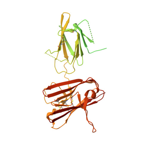
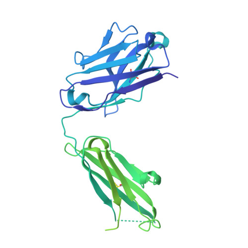
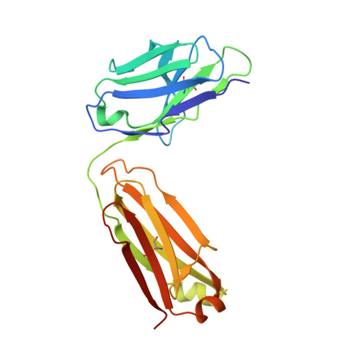
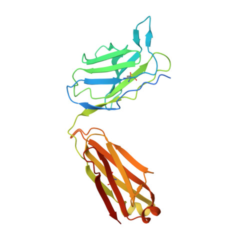
Structure of the malaria vaccine candidate Pfs48/45 and its recognition by transmission blocking antibodies.

Ko, K.T.Lennartz, F.Mekhaiel, D.Guloglu, B.Marini, A.Deuker, D.J.Long, C.A.Jore, M.M.Miura, K.Biswas, S.Higgins, M.K.

(2022) Nat Commun 13: 5603-5603

  • DOI: https://doi.org/10.1038/s41467-022-33379-6
  • Primary Citation of Related Structures:  
    7ZWF, 7ZWI, 7ZWM, 7ZXF, 7ZXG

  • PubMed Abstract: 

    An effective malaria vaccine remains a global health priority and vaccine immunogens which prevent transmission of the parasite will have important roles in multi-component vaccines. One of the most promising candidates for inclusion in a transmission-blocking malaria vaccine is the gamete surface protein Pfs48/45, which is essential for development of the parasite in the mosquito midgut. Indeed, antibodies which bind Pfs48/45 can prevent transmission if ingested with the parasite as part of the mosquito bloodmeal. Here we present the structure of full-length Pfs48/45, showing its three domains to form a dynamic, planar, triangular arrangement. We reveal where transmission-blocking and non-blocking antibodies bind on Pfs48/45. Finally, we demonstrate that antibodies which bind across this molecule can be transmission-blocking. These studies will guide the development of future Pfs48/45-based vaccine immunogens.


  • Organizational Affiliation

    Department of Biochemistry, South Parks Road, University of Oxford, Oxford, OX1 3QU, UK.


Macromolecules
Find similar proteins by:  (by identity cutoff)  |  3D Structure
Entity ID: 1
MoleculeChains Sequence LengthOrganismDetailsImage
Gametocyte surface protein P45/48
A, F
428Plasmodium falciparumMutation(s): 0 
Gene Names: PF45/48PFS45-48PFS45/48PF13_0247PF3D7_1346700
UniProt
Find proteins for Q8I6T1 (Plasmodium falciparum (isolate 3D7))
Explore Q8I6T1 
Go to UniProtKB:  Q8I6T1
Entity Groups  
Sequence Clusters30% Identity50% Identity70% Identity90% Identity95% Identity100% Identity
UniProt GroupQ8I6T1
Glycosylation
Glycosylation Sites: 2
Sequence Annotations
Expand
  • Reference Sequence
Find similar proteins by:  (by identity cutoff)  |  3D Structure
Entity ID: 2
MoleculeChains Sequence LengthOrganismDetailsImage
32F3 heavy chain
B, G
444Mus musculusMutation(s): 0 
Entity Groups  
Sequence Clusters30% Identity50% Identity70% Identity90% Identity95% Identity100% Identity
Sequence Annotations
Expand
  • Reference Sequence
Find similar proteins by:  (by identity cutoff)  |  3D Structure
Entity ID: 3
MoleculeChains Sequence LengthOrganismDetailsImage
32F3 light chain
C, H
213Mus musculusMutation(s): 0 
Entity Groups  
Sequence Clusters30% Identity50% Identity70% Identity90% Identity95% Identity100% Identity
Sequence Annotations
Expand
  • Reference Sequence
Find similar proteins by:  (by identity cutoff)  |  3D Structure
Entity ID: 4
MoleculeChains Sequence LengthOrganismDetailsImage
10D8 heavy chain
D, I
466Mus musculusMutation(s): 0 
Entity Groups  
Sequence Clusters30% Identity50% Identity70% Identity90% Identity95% Identity100% Identity
Sequence Annotations
Expand
  • Reference Sequence
Find similar proteins by:  (by identity cutoff)  |  3D Structure
Entity ID: 5
MoleculeChains Sequence LengthOrganismDetailsImage
10D8 light chain
E, J
240Mus musculusMutation(s): 0 
Entity Groups  
Sequence Clusters30% Identity50% Identity70% Identity90% Identity95% Identity100% Identity
Sequence Annotations
Expand
  • Reference Sequence
Oligosaccharides

Help

Entity ID: 6
MoleculeChains Length2D Diagram Glycosylation3D Interactions
beta-D-mannopyranose-(1-4)-2-acetamido-2-deoxy-beta-D-glucopyranose-(1-4)-2-acetamido-2-deoxy-beta-D-glucopyranose
K
3N-Glycosylation
Glycosylation Resources
GlyTouCan:  G15407YE
GlyCosmos:  G15407YE
GlyGen:  G15407YE
Entity ID: 7
MoleculeChains Length2D Diagram Glycosylation3D Interactions
beta-D-mannopyranose-(1-4)-2-acetamido-2-deoxy-beta-D-glucopyranose-(1-4)-[alpha-L-fucopyranose-(1-6)]2-acetamido-2-deoxy-beta-D-glucopyranose
L
4N-Glycosylation
Glycosylation Resources
GlyTouCan:  G32152BH
GlyCosmos:  G32152BH
GlyGen:  G32152BH
Experimental Data & Validation

Experimental Data

  • Method: X-RAY DIFFRACTION
  • Resolution: 3.69 Å
  • R-Value Free: 0.280 
  • R-Value Work: 0.255 
  • R-Value Observed: 0.257 
  • Space Group: P 21 21 21
Unit Cell:
Length ( Å )Angle ( ˚ )
a = 108.583α = 90
b = 158.451β = 90
c = 186.163γ = 90
Software Package:
Software NamePurpose
BUSTERrefinement
PHENIXrefinement
xia2data reduction
xia2data scaling
PHASERphasing

Structure Validation

View Full Validation Report


Ligand Structure Quality Assessment 


Entry History & Funding Information

Deposition Data


Funding OrganizationLocationGrant Number
Medical Research Council (MRC, United Kingdom)United KingdomMR/R001138/1
Wellcome TrustUnited Kingdom218482/Z/19/Z
Wellcome TrustUnited Kingdom101020/Z/13/Z

Revision History  (Full details and data files)

  • Version 1.0: 2022-06-22
    Type: Initial release
  • Version 1.1: 2022-10-12
    Changes: Database references
  • Version 1.2: 2024-01-31
    Changes: Data collection, Refinement description