7ZVX

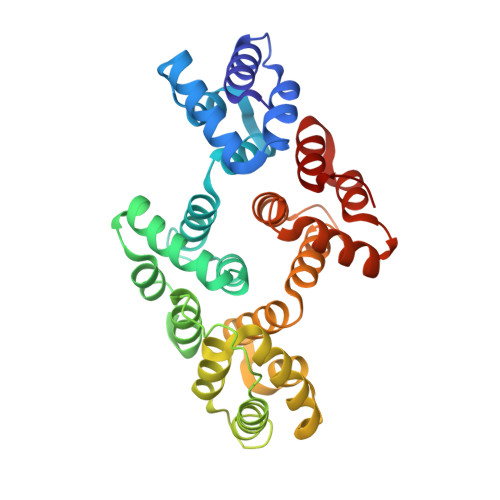
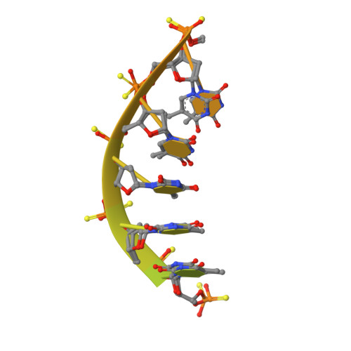
Crystal structure of human Annexin A2 in complex with full phosphorothioate 5-10 2'-methoxyethyl DNA gapmer antisense oligonucleotide solved at 2.4 A resolution


Experimental Data Snapshot

  • Method: X-RAY DIFFRACTION
  • Resolution: 2.40 Å
  • R-Value Free: 0.242 
  • R-Value Work: 0.185 
  • R-Value Observed: 0.188 

wwPDB Validation   3D Report Full Report


This is version 1.3 of the entry. See complete history


Literature

Structures of annexin A2-PS DNA complexes show dominance of hydrophobic interactions in phosphorothioate binding.

Hyjek-Skladanowska, M.Anderson, B.A.Mykhaylyk, V.Orr, C.Wagner, A.Poznanski, J.T.Skowronek, K.Seth, P.Nowotny, M.

(2023) Nucleic Acids Res 51: 1409-1423

  • DOI: https://doi.org/10.1093/nar/gkac774
  • Primary Citation of Related Structures:  
    7ZVN, 7ZVX

  • PubMed Abstract: 

    The introduction of phosphorothioate (PS) linkages to the backbone of therapeutic nucleic acids substantially increases their stability and potency. It also affects their interactions with cellular proteins, but the molecular mechanisms that underlie this effect are poorly understood. Here, we report structural and biochemical studies of interactions between annexin A2, a protein that does not possess any known canonical DNA binding domains, and phosphorothioate-modified antisense oligonucleotides. We show that a unique mode of hydrophobic interactions between a sulfur atom of the phosphorothioate group and lysine and arginine residues account for the enhanced affinity of modified nucleic acid for the protein. Our results demonstrate that this mechanism of interaction is observed not only for nucleic acid-binding proteins but can also account for the association of PS oligonucleotides with other proteins. Using the anomalous diffraction of sulfur, we showed that preference for phosphorothioate stereoisomers is determined by the hydrophobic environment around the PS linkage that comes not only from protein but also from additional structural features within the ASO such as 5-Me groups on cytosine nucleobases.


  • Organizational Affiliation

    Laboratory of Protein Structure, International Institute of Molecular and Cell Biology, Warsaw 02-109, Poland.


Macromolecules

Find similar proteins by:  (by identity cutoff)  |  3D Structure
Entity ID: 1
MoleculeChains Sequence LengthOrganismDetailsImage
Annexin A2
A, B
307Homo sapiensMutation(s): 1 
Gene Names: ANXA2ANX2ANX2L4CAL1HLPC2D
UniProt & NIH Common Fund Data Resources
Find proteins for P07355 (Homo sapiens)
Explore P07355 
Go to UniProtKB:  P07355
PHAROS:  P07355
GTEx:  ENSG00000182718 
Entity Groups  
Sequence Clusters30% Identity50% Identity70% Identity90% Identity95% Identity100% Identity
UniProt GroupP07355
Sequence Annotations
Expand
  • Reference Sequence
Find similar nucleic acids by:  (by identity cutoff)  |  3D Structure
Entity ID: 2
MoleculeChains LengthOrganismImage
2'-methoxyethyl DNA gapmer antisense oligonucleotide
C, D
30synthetic construct
Sequence Annotations
Expand
  • Reference Sequence
Small Molecules
Ligands 2 Unique
IDChains Name / Formula / InChI Key2D Diagram3D Interactions
EDO
Query on EDO

Download Ideal Coordinates CCD File 
AA [auth B]
BA [auth B]
J [auth A]
K [auth A]
L [auth A]
AA [auth B],
BA [auth B],
J [auth A],
K [auth A],
L [auth A],
M [auth A],
N [auth A],
O [auth A],
P [auth A],
W [auth B],
X [auth B],
Y [auth B],
Z [auth B]
1,2-ETHANEDIOL
C2 H6 O2
LYCAIKOWRPUZTN-UHFFFAOYSA-N
CA
Query on CA

Download Ideal Coordinates CCD File 
E [auth A]
F [auth A]
G [auth A]
H [auth A]
I [auth A]
E [auth A],
F [auth A],
G [auth A],
H [auth A],
I [auth A],
Q [auth B],
R [auth B],
S [auth B],
T [auth B],
U [auth B],
V [auth B]
CALCIUM ION
Ca
BHPQYMZQTOCNFJ-UHFFFAOYSA-N
Experimental Data & Validation

Experimental Data

  • Method: X-RAY DIFFRACTION
  • Resolution: 2.40 Å
  • R-Value Free: 0.242 
  • R-Value Work: 0.185 
  • R-Value Observed: 0.188 
  • Space Group: P 1 21 1
Unit Cell:
Length ( Å )Angle ( ˚ )
a = 50.23α = 90
b = 60.36β = 97.17
c = 128.43γ = 90
Software Package:
Software NamePurpose
PHENIXrefinement
XDSdata reduction
XSCALEdata scaling
PHASERphasing

Structure Validation

View Full Validation Report


Entry History & Funding Information

Deposition Data


Funding OrganizationLocationGrant Number
Not funded--

Revision History  (Full details and data files)

  • Version 1.0: 2022-09-14
    Type: Initial release
  • Version 1.1: 2022-10-05
    Changes: Database references
  • Version 1.2: 2023-03-01
    Changes: Database references
  • Version 1.3: 2024-02-07
    Changes: Data collection, Refinement description