7ZVS | pdb_00007zvs

Crystal structure of the Schistosoma mansoni VKR2 kinase domain in an active-like state (ADP-bound)


Experimental Data Snapshot

  • Method: X-RAY DIFFRACTION
  • Resolution: 3.07 Å
  • R-Value Free: 
    0.252 (Depositor), 0.257 (DCC) 
  • R-Value Work: 
    0.217 (Depositor), 0.221 (DCC) 
  • R-Value Observed: 
    0.219 (Depositor) 

Starting Model: experimental
View more details

wwPDB Validation 3D Report Full Report

Validation slider image for 7ZVS

Ligand Structure Quality Assessment 


This is version 1.3 of the entry. See complete history

Literature

Identification of Inhibitors of the Schistosoma mansoni VKR2 Kinase Domain.

Mathavan, I.Liu, L.J.Robinson, S.W.El-Sakkary, N.Elatico, A.J.J.Gomez, D.Nellas, R.Owens, R.J.Zuercher, W.Navratilova, I.Caffrey, C.R.Beis, K.

(2022) ACS Med Chem Lett 13: 1715-1722

  • DOI: https://doi.org/10.1021/acsmedchemlett.2c00248
  • Primary Citation Related Structures: 
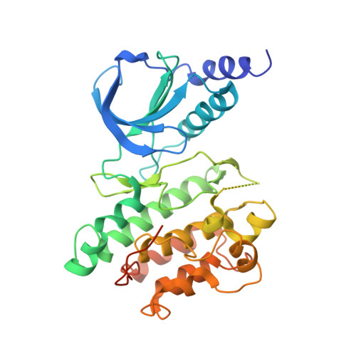
    7ZVS

  • PubMed Abstract: 

    Schistosomiasis is a neglected tropical disease caused by parasitic flatworms. Current treatment relies on just one partially effective drug, praziquantel (PZQ). Schistosoma mansoni Venus Kinase Receptors 1 and 2 (SmVKR1 and SmVKR2) are important for parasite growth and egg production, and are potential targets for combating schistosomiasis. VKRs consist of an extracellular Venus Flytrap Module (VFTM) linked via a transmembrane helix to a kinase domain. Here, we initiated a drug discovery effort to inhibit the activity of the SmVKR2 kinase domain (SmVKR2 KD ) by screening the GSK published kinase inhibitor set 2 (PKIS2). We identified several inhibitors, of which four were able to inhibit its enzymatic activity and induced phenotypic changes in ex vivo S. mansoni . Our crystal structure of the SmVKR2 KD displays an active-like state that sheds light on the activation process of VKRs. Our data provide a basis for the further exploration of SmVKR2 as a possible drug target.


  • Organizational Affiliation
    • Department of Life Sciences, Imperial College London, Exhibition Road, London, South Kensington SW7 2AZ, United Kingdom.

Macromolecule Content 

  • Total Structure Weight: 117.27 kDa 
  • Atom Count: 7,186 
  • Modeled Residue Count: 896 
  • Deposited Residue Count: 1,023 
  • Unique protein chains: 1

Macromolecules

Find similar proteins by:|  3D Structure
Entity ID: 1
MoleculeChains  Sequence LengthOrganismDetailsImage
Venus kinase receptor 2
A, B, C
341Schistosoma mansoniMutation(s): 0 
UniProt
Find proteins for A0A3Q0KQ92 (Schistosoma mansoni)
Explore A0A3Q0KQ92 
Go to UniProtKB:  A0A3Q0KQ92
Entity Groups
Sequence Clusters30% Identity50% Identity70% Identity90% Identity95% Identity100% Identity
UniProt GroupA0A3Q0KQ92
Sequence Annotations
Expand
Reference Sequence

Experimental Data & Validation

Experimental Data

  • Method: X-RAY DIFFRACTION
  • Resolution: 3.07 Å
  • R-Value Free:  0.252 (Depositor), 0.257 (DCC) 
  • R-Value Work:  0.217 (Depositor), 0.221 (DCC) 
  • R-Value Observed: 0.219 (Depositor) 
Space Group: C 1 2 1
Unit Cell:
Length ( Å )Angle ( ˚ )
a = 206.258α = 90
b = 97.239β = 97.52
c = 72.944γ = 90
Software Package:
Software NamePurpose
PHENIXrefinement
autoPROCdata reduction
autoPROCdata scaling
PHASERphasing

Structure Validation

View Full Validation Report


Ligand Structure Quality Assessment 


Entry History 

& Funding Information

Deposition Data


Funding OrganizationLocationGrant Number
Medical Research Council (MRC, United Kingdom)United KingdomMR/N019113/1

Revision History  (Full details and data files)

  • Version 1.0: 2022-10-12
    Type: Initial release
  • Version 1.1: 2022-10-26
    Changes: Database references
  • Version 1.2: 2022-11-30
    Changes: Database references
  • Version 1.3: 2024-01-31
    Changes: Data collection, Refinement description